Copyright
©The Author(s) 2024.
World J Clin Cases. Jun 6, 2024; 12(16): 2813-2821
Published online Jun 6, 2024. doi: 10.12998/wjcc.v12.i16.2813
Published online Jun 6, 2024. doi: 10.12998/wjcc.v12.i16.2813
Figure 1 The whole exon gene test of the patient with Williams-Beuren syndrome.
The Whole Exome Gene Detection V6 revealed a copy number deletion of approximately 1.49Mb in the long arm of chromosome 7 (7q11.23), which includes the haploinsufficient gene ELN.
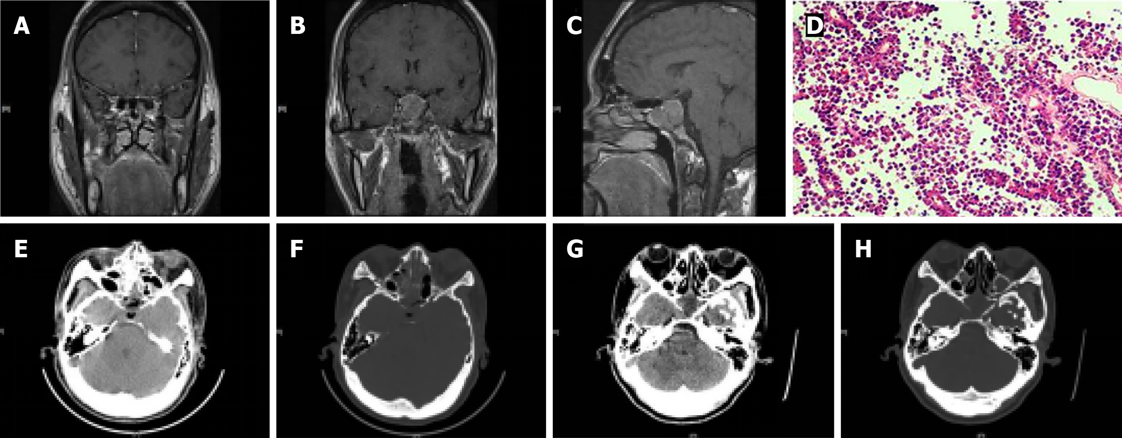
Figure 2 Preoperative and postoperative changes and histopathology of the patient with acromegaly.
A: The magnetic resonance scan of the pituitary indicated bilateral inferior turbinate hypertrophy; B and C: The coronal and sagittal positions revealed neoplastic lesions in the pituitary region, with pituitary tumors invading the bilateral sphenoid sinuses; D: Pathology confirmed the presence of a pituitary adenoma; E and F: The day after surgery, a pituitary computed tomography (CT) scan showed enlargement of the sellar region, with no residual tumor tissue observed. Additionally, there was visible liquid signal filling; G: On the 9th postoperative day, a small amount of gas was observed in the soft tissue window of the pituitary CT; H: The bone window revealed further enlargement of the sellar region.
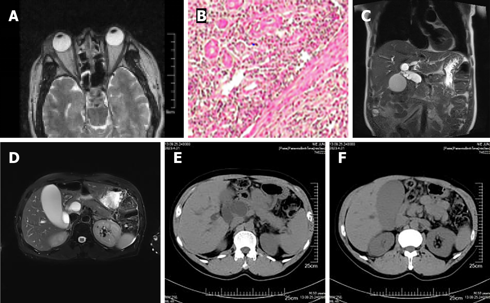
Figure 3 The imaging and histopathological examination of some organs and tissues of the patient with IgG4-related diseases.
A: Orbital magnetic resonance (MR) showed space-occupying lesions in the lacrimal gland of the right eye; B: Histopathology of the eye revealed an inflammatory pseudotumor of the lacrimal gland; C: Epigastric MR indicated dilatation of the common bile duct, stenosis of the lower common bile duct; D: Enlargement of the gallbladder, cholestasis, and enlargement of the head of the pancreas; E: Epigastric computed tomography showed significant dilation of the common bile duct; F: Enlargement of the gallbladder and cholestasis.
- Citation: Song WR, Xu XH, Li J, Yu J, Li YX. Secondary diabetes due to different etiologies: Four case reports. World J Clin Cases 2024; 12(16): 2813-2821
- URL: https://www.wjgnet.com/2307-8960/full/v12/i16/2813.htm
- DOI: https://dx.doi.org/10.12998/wjcc.v12.i16.2813


