Copyright
©The Author(s) 2024.
World J Clin Cases. May 26, 2024; 12(15): 2678-2681
Published online May 26, 2024. doi: 10.12998/wjcc.v12.i15.2678
Published online May 26, 2024. doi: 10.12998/wjcc.v12.i15.2678
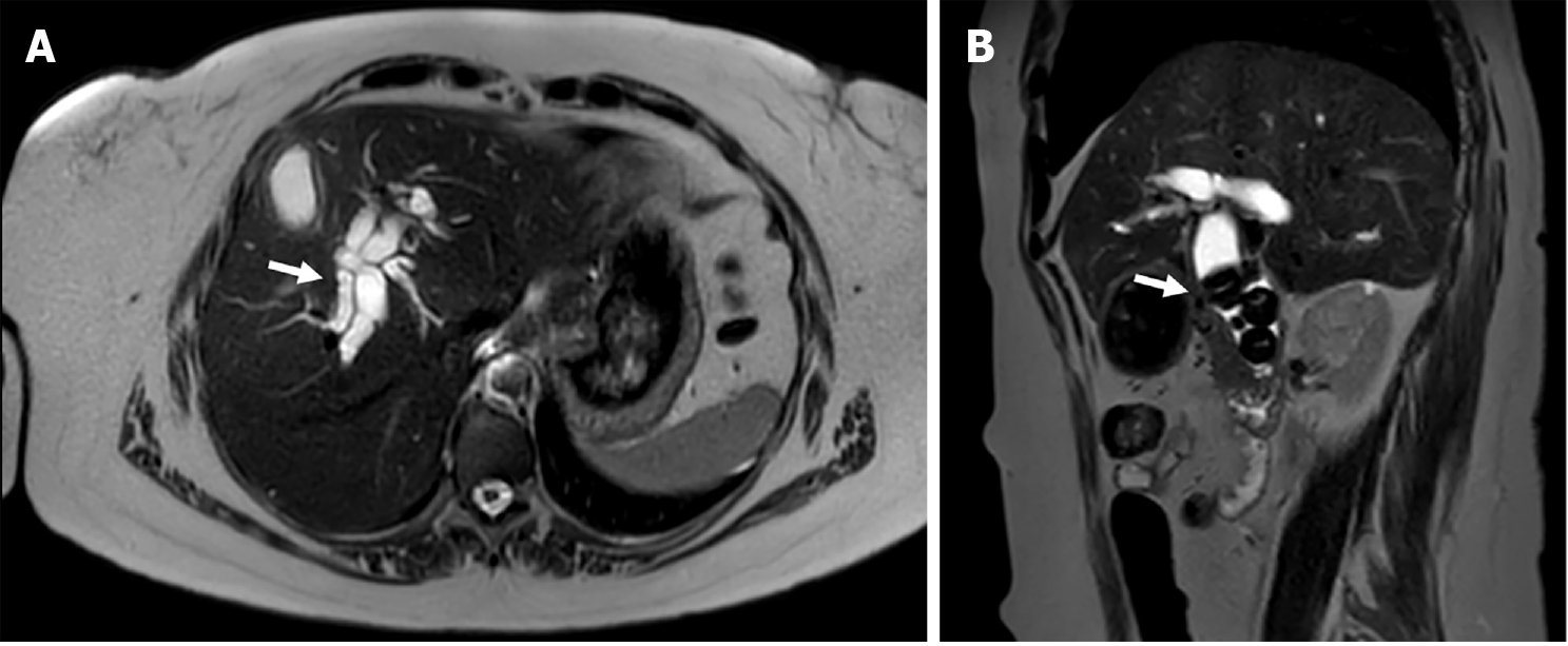
Figure 1 Distal choledocholithiasis in a 53-year-old man with acute secondary cholangitis.
A: Axial magnetic resonance T2-weighted images depicts intra- and extrahepatic biliary ductal dilatation; B: Multiple void signal images within the lumen of the common bile duct.
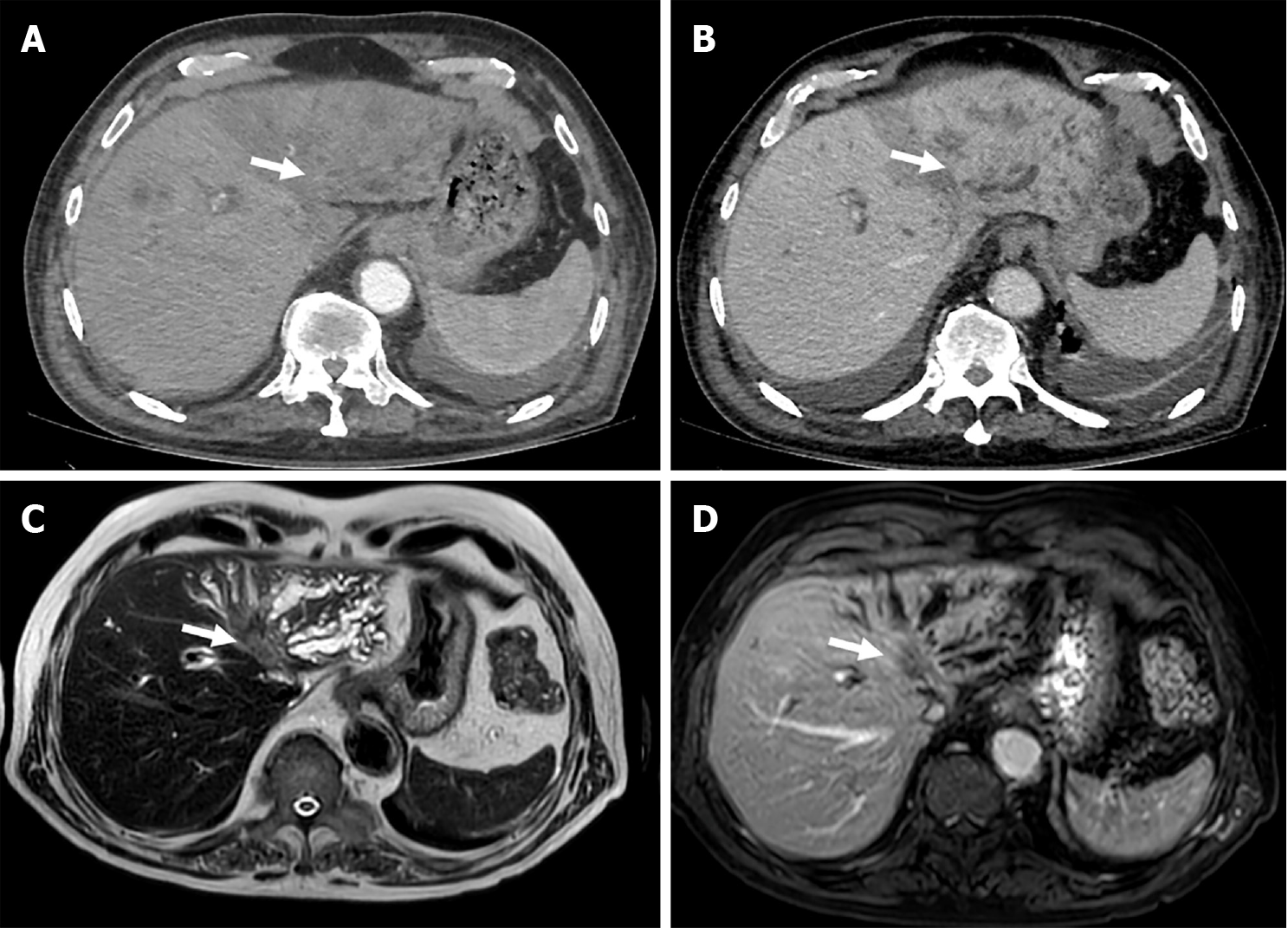
Figure 2 69-years-old woman with biliary obstruction secondary to intrahepatic mass-forming type cholangiocarcinoma with tumoral involvement of the biliary ducts confluence.
A: Axial late computed tomography arterial phase depicts a soft-tissue mass in left hepatic lobe, with delayed phase enhancement; B: Dilatation of intrahepatic ducts; C: Axial magnetic resonance images demonstrate a T2-weighted images slightly hyperintense; D: T1-weighted images hypointense heterogeneous mass occluding the confluence of the hepatic ducts with moderate dilatation of left lobar intrahepatic bile ducts, and tumoral involvement of the left intrahepatic biliary branches.
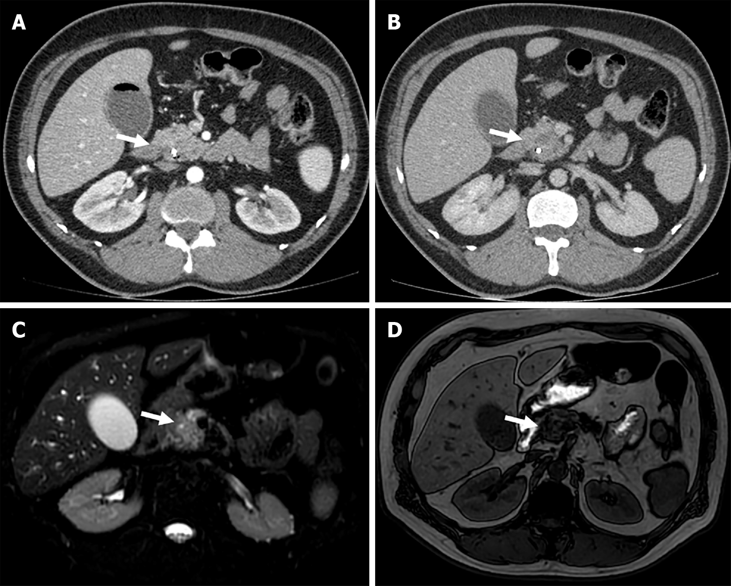
Figure 3 Pancreatic ductal adenocarcinoma in a 72-years-old man with obstructive jaundice.
A: Axial contrast-enhanced computed tomography image shows a well-circumscribed exophytic tumor in the pancreatic head with heterogeneuos enhancement in delayed arterial; B: Portovenous phase; C: Axial magnetic resonance fat suppression T2-weighted images image showing a mildly hyperdense irregular mass in the head of the pancreas; D: T1-weighted images with hypointense signal.
- Citation: Lindner C. Imaging features of malignant vs stone-induced biliary obstruction: Aspects to consider. World J Clin Cases 2024; 12(15): 2678-2681
- URL: https://www.wjgnet.com/2307-8960/full/v12/i15/2678.htm
- DOI: https://dx.doi.org/10.12998/wjcc.v12.i15.2678


