Copyright
©The Author(s) 2024.
World J Clin Cases. May 26, 2024; 12(15): 2664-2671
Published online May 26, 2024. doi: 10.12998/wjcc.v12.i15.2664
Published online May 26, 2024. doi: 10.12998/wjcc.v12.i15.2664
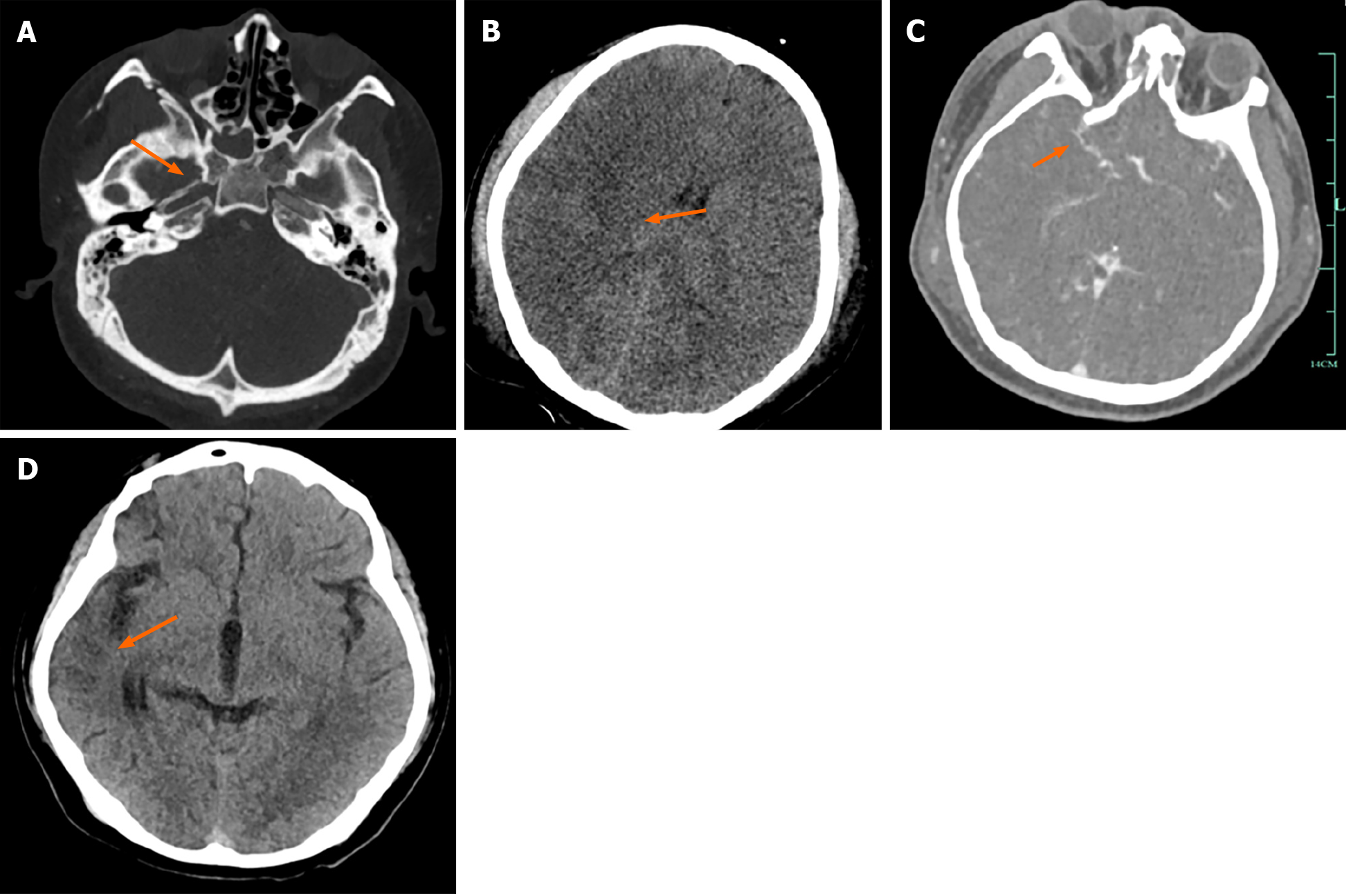
Figure 1 Cranial imaging scans of Case 1.
A: A sphenoid bone fracture and non-opacification of the right internal carotid artery (indicated by an arrow); B: Ischemia in the right cerebral hemisphere (arrow), with no midline shift; C: Compensation by intracranial vascular collateral circulation (arrow); D: Demonstrates a reduction in the right-sided ischemic area after treatment and recovery (arrow).
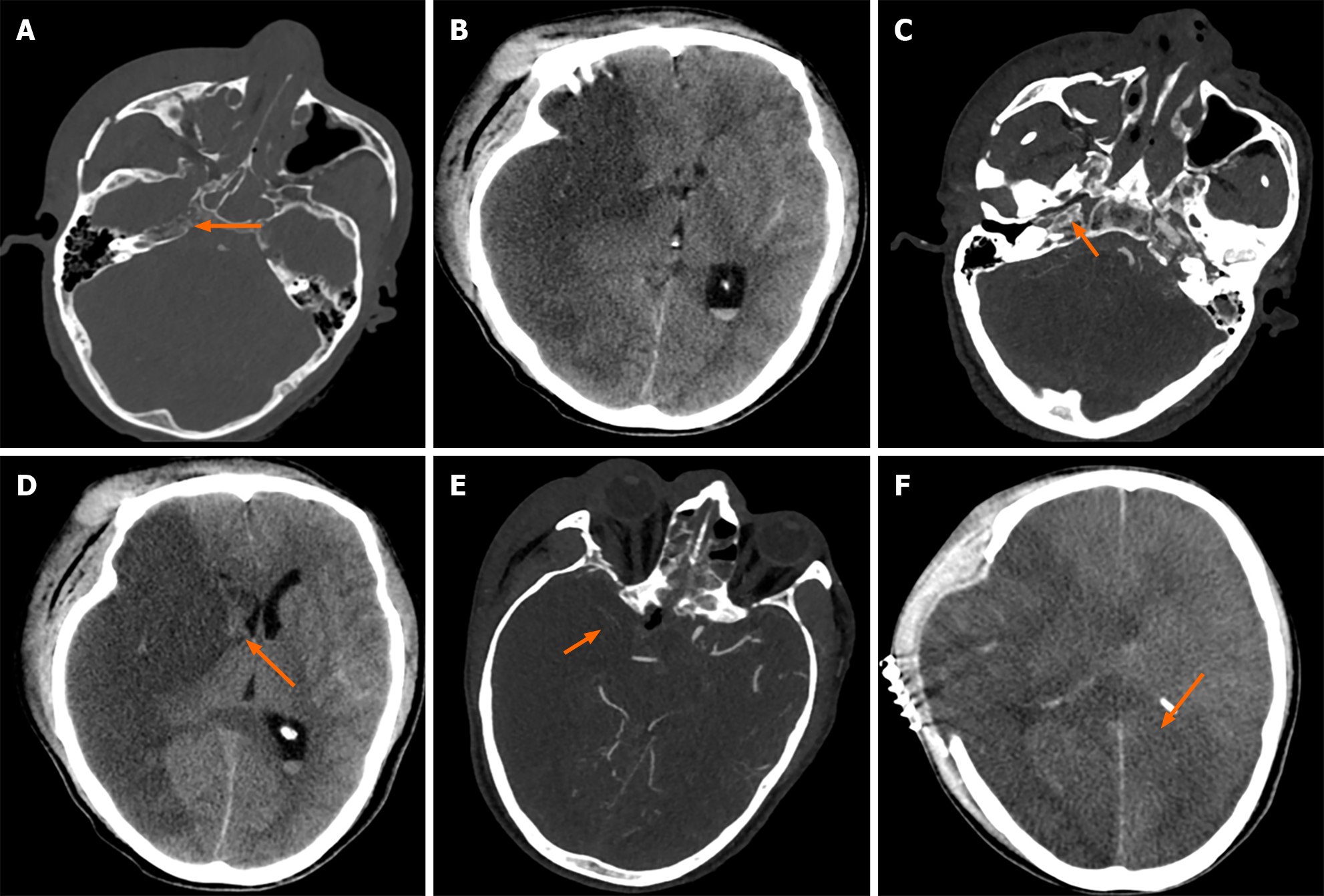
Figure 2 Cranial imaging scans of Case 2.
A: A fracture of the right temporal bone's petrous part (arrow), with an incomplete structure of the carotid canal; B: Ischemic lesions in the right cerebral hemisphere; C: Occlusion of the right internal carotid artery (arrow); D: Ischemia in the right cerebral hemisphere (arrow), with midline shift and brain herniation; E: Poor compensation of intracranial vascular collateral circulation (arrow); F: The area of cerebral ischemia has increased following treatment (arrow), suggesting a poor prognosis.
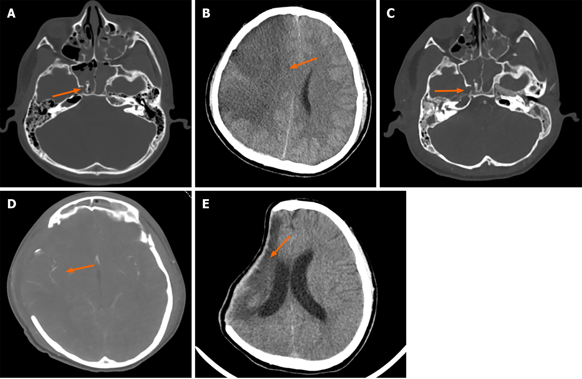
Figure 3 Cranial imaging scans of Case 3.
A: A right sphenoid bone fracture (arrow), B: Right cerebral ischemia (arrow), midline shift, brain herniation; C: Right internal carotid artery occlusion (arrow); D: Compensation through collateral circulation in the right brain tissue (arrow); E: Post-treatment recovery with atrophy of the right ischemic area, slightly reduced in size.
- Citation: Shangguan PX, Zhou KC. Imaging characteristics and treatment strategies for carotid artery occlusion caused by skull base fracture: Three case reports. World J Clin Cases 2024; 12(15): 2664-2671
- URL: https://www.wjgnet.com/2307-8960/full/v12/i15/2664.htm
- DOI: https://dx.doi.org/10.12998/wjcc.v12.i15.2664


