Copyright
©The Author(s) 2024.
World J Clin Cases. May 26, 2024; 12(15): 2529-2541
Published online May 26, 2024. doi: 10.12998/wjcc.v12.i15.2529
Published online May 26, 2024. doi: 10.12998/wjcc.v12.i15.2529
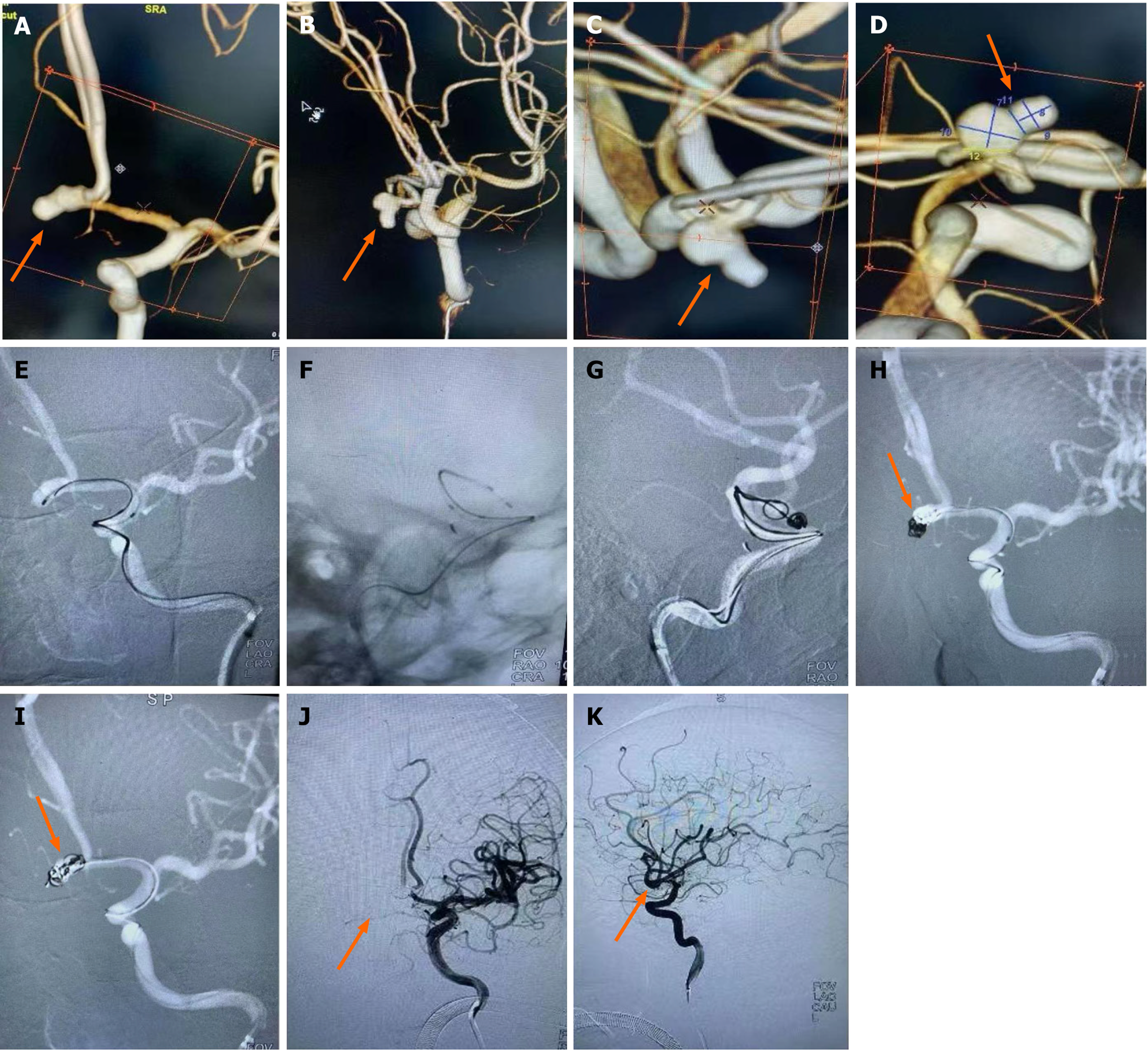
Figure 1 Imaging data of endovascular treatment of typical double-lobed anterior communicating artery aneurysms.
A-D: Double-lobed anterior communicating artery aneurysms, the lobes were similar in size to the mother aneurysm and were not on the same long axis (arrows); E-I: Double microcatheters were placed at different working angles to different lobes, and each lobe was embolized alternately in the double microtubes (arrows); J and K: After embolization, the aneurysm was not visualized and bilateral A2 was unobstructed (arrows).
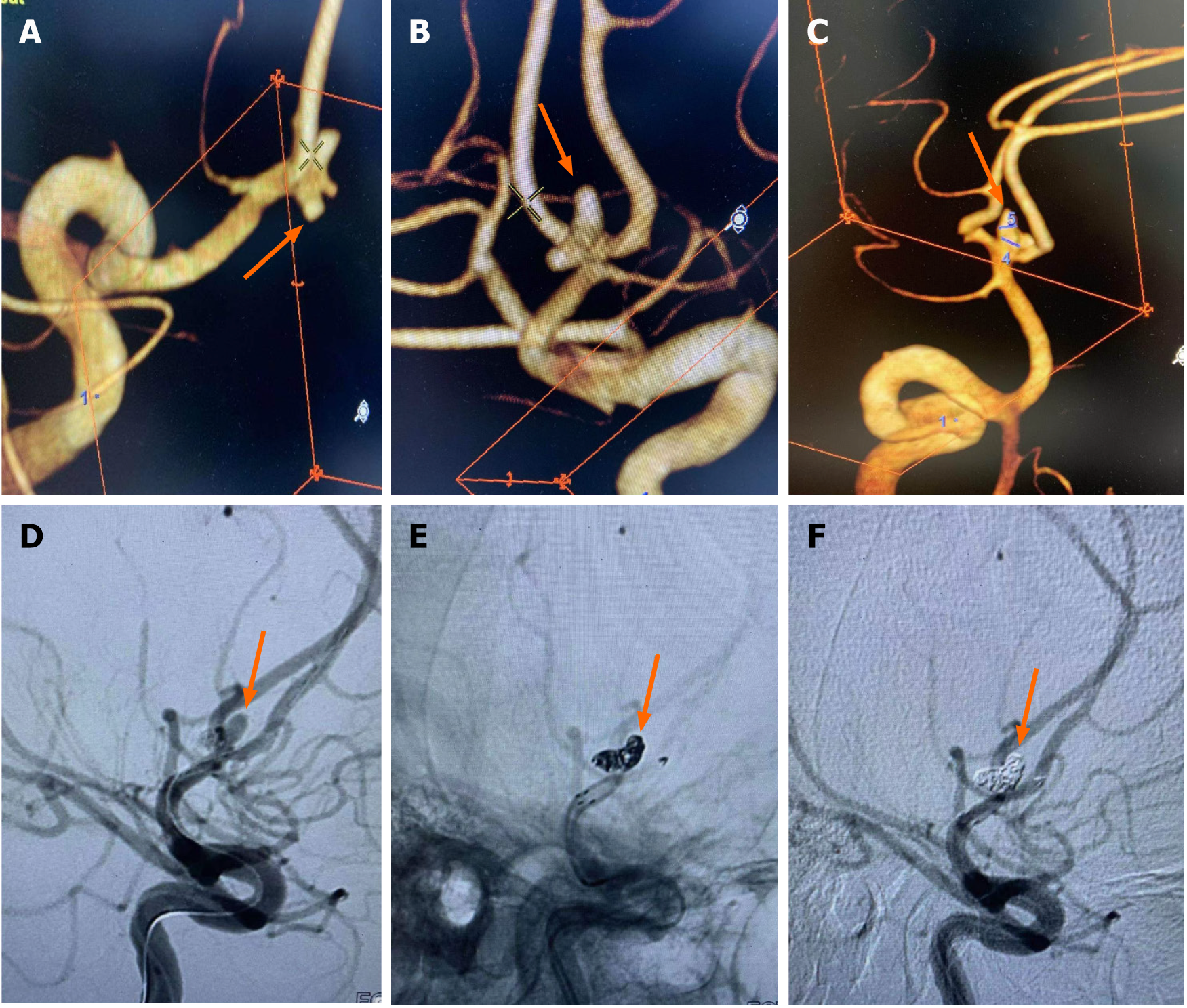
Figure 2 Imaging data of endovascular treatment of typical anterior communicating artery trilobular aneurysms.
A-C: Anterior communicating artery trilobular aneurysms (arrows); D: First embolization of the posterior lobe, completely release the stent, embolization of the microcatheter through the stent mesh to the forward lobe, and continue to embolization of the aneurysm (arrow); E-F: Complete embolization of the aneurysm, bilateral A2 patency (arrows).
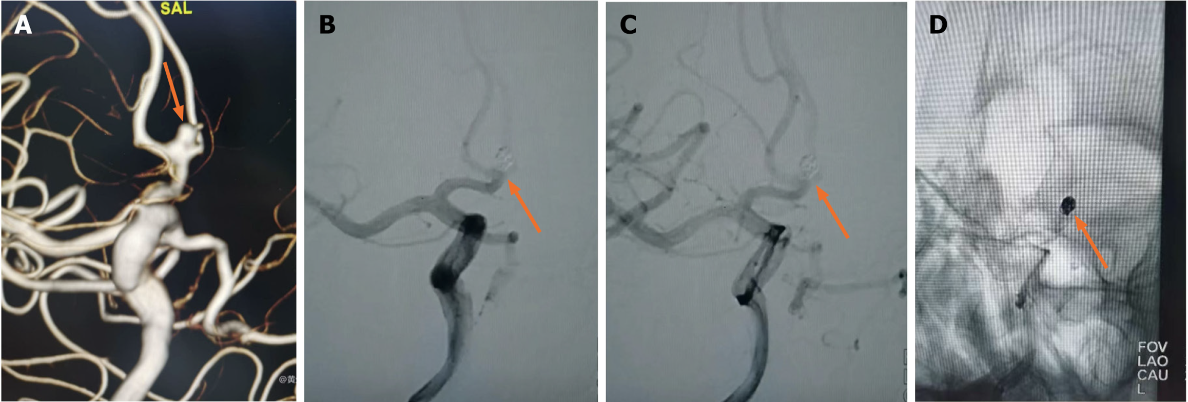
Figure 3 Imaging data of single microcatheter embolization of anterior communicating aneurysm.
A: The top of the anterior communicating aneurysm was accompanied by a subaneurysm (less than 1mm), which was on the same long axis as the mother aneurysm (arrow); B-D: Single microcatheter embolization of the aneurysm, without deliberately treating the subaneurysm, after dense embolization, the aneurysm did not develop, and bilateral A2 was unobstructed (arrows).
Figure 4 Imaging data of communicating artery aneurysms before embolization with double microcatheter.
A: The top of anterior communicating aneurysm is accompanied by A daughter tumor (larger than 1 mm), and the daughter tumor and the mother tumor are on the same long axis (arrow); B: Double microcatheter embolization of aneurysm, alternate filling of daughter tumor and mother tumor (arrow); C and D: Dense embolization, the aneurysm did not develop, and bilateral A2 was patency (arrows).
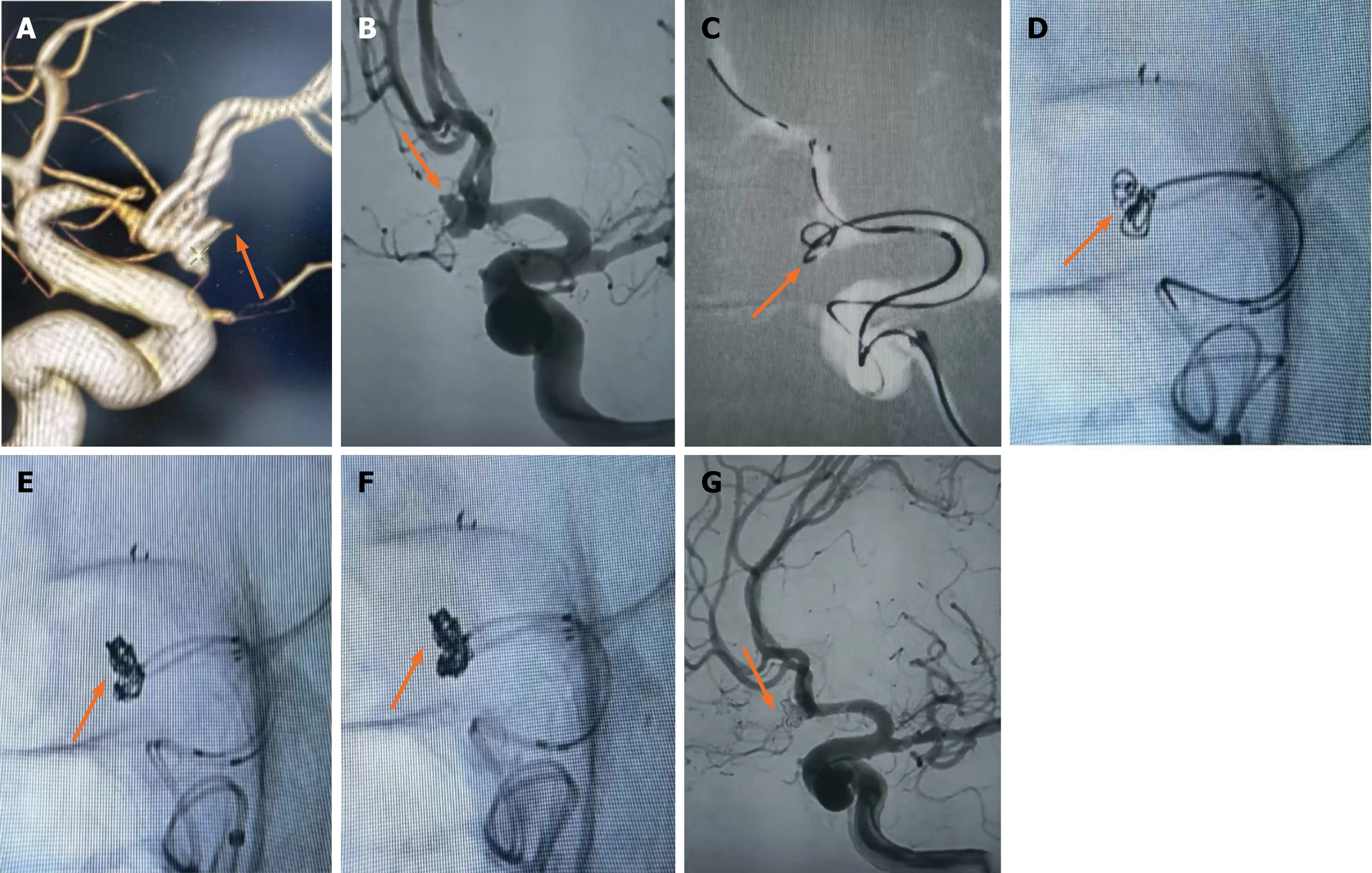
Figure 5 Imaging data of communicating artery aneurysm before stent assisted embolization.
A: Anterior communicating double-lobular aneurysm with A1 segment spasm (arrow); B: Segment A1 spasm improved after continuous nimodipine pumping treatment (arrow); C: In the semi-release state of the support, release the spring ring several times in the pointing down lobe (arrow); D-F: Completely released the stent, embolized the microcatheter through the stent mesh into the other lobe, the double microcatheter alternately into the spring coil (arrows); G: After the completion of embolization, the aneurysm did not develop and bilateral A2 was unobstructed (arrow).
- Citation: Huang SX, Ai XP, Kang ZH, Chen ZY, Li RM, Wu ZC, Zhu F. Endovascular treatment of ruptured lobulated anterior communicating artery aneurysms: A retrospective study of 24 patients. World J Clin Cases 2024; 12(15): 2529-2541
- URL: https://www.wjgnet.com/2307-8960/full/v12/i15/2529.htm
- DOI: https://dx.doi.org/10.12998/wjcc.v12.i15.2529


