Copyright
©The Author(s) 2024.
World J Clin Cases. May 16, 2024; 12(14): 2457-2462
Published online May 16, 2024. doi: 10.12998/wjcc.v12.i14.2457
Published online May 16, 2024. doi: 10.12998/wjcc.v12.i14.2457
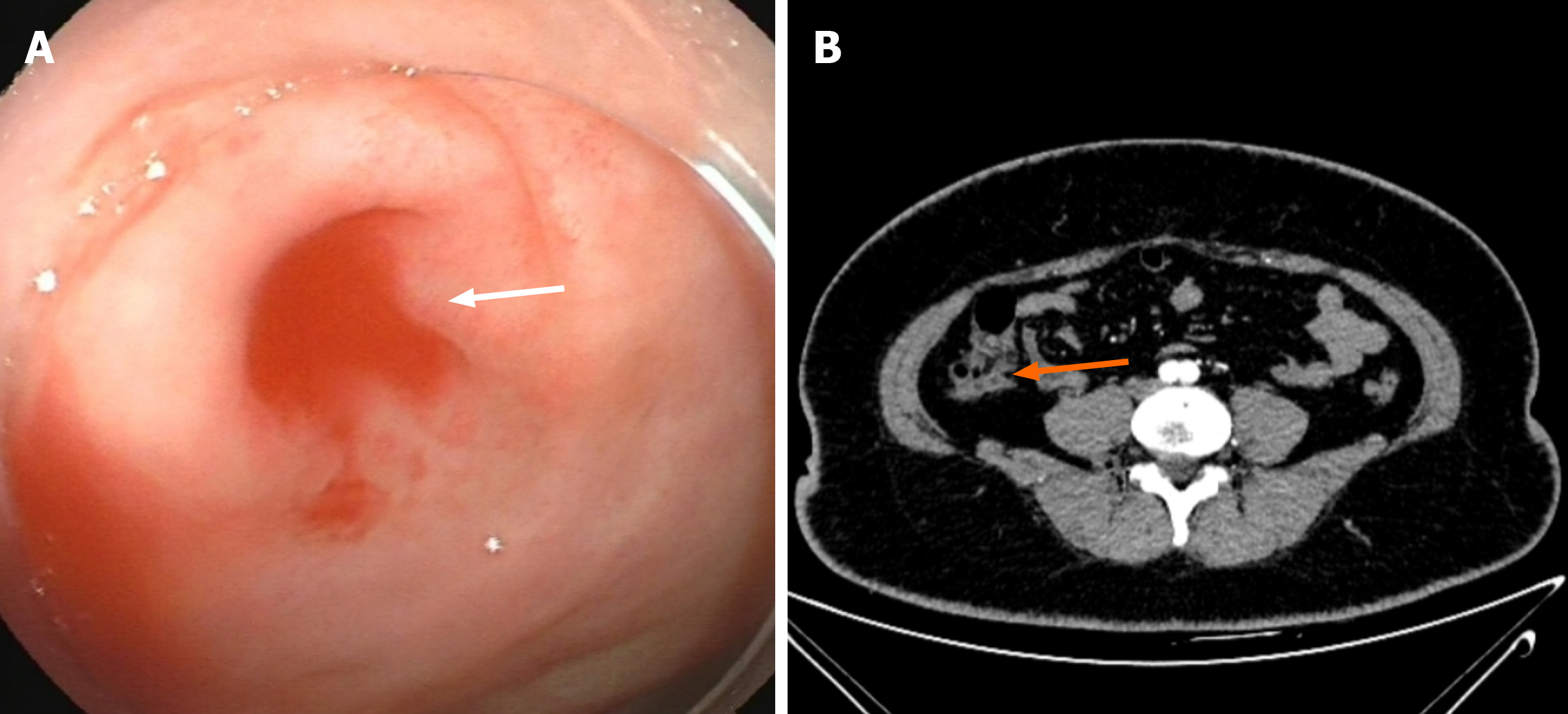
Figure 1 Colonoscopy and contrast-enhanced abdominal computed tomography scan.
A: Continuous bleeding was observed at the orifice of the appendix; B: Structural disorder in the ileocecal area, and the appendix was not clear.
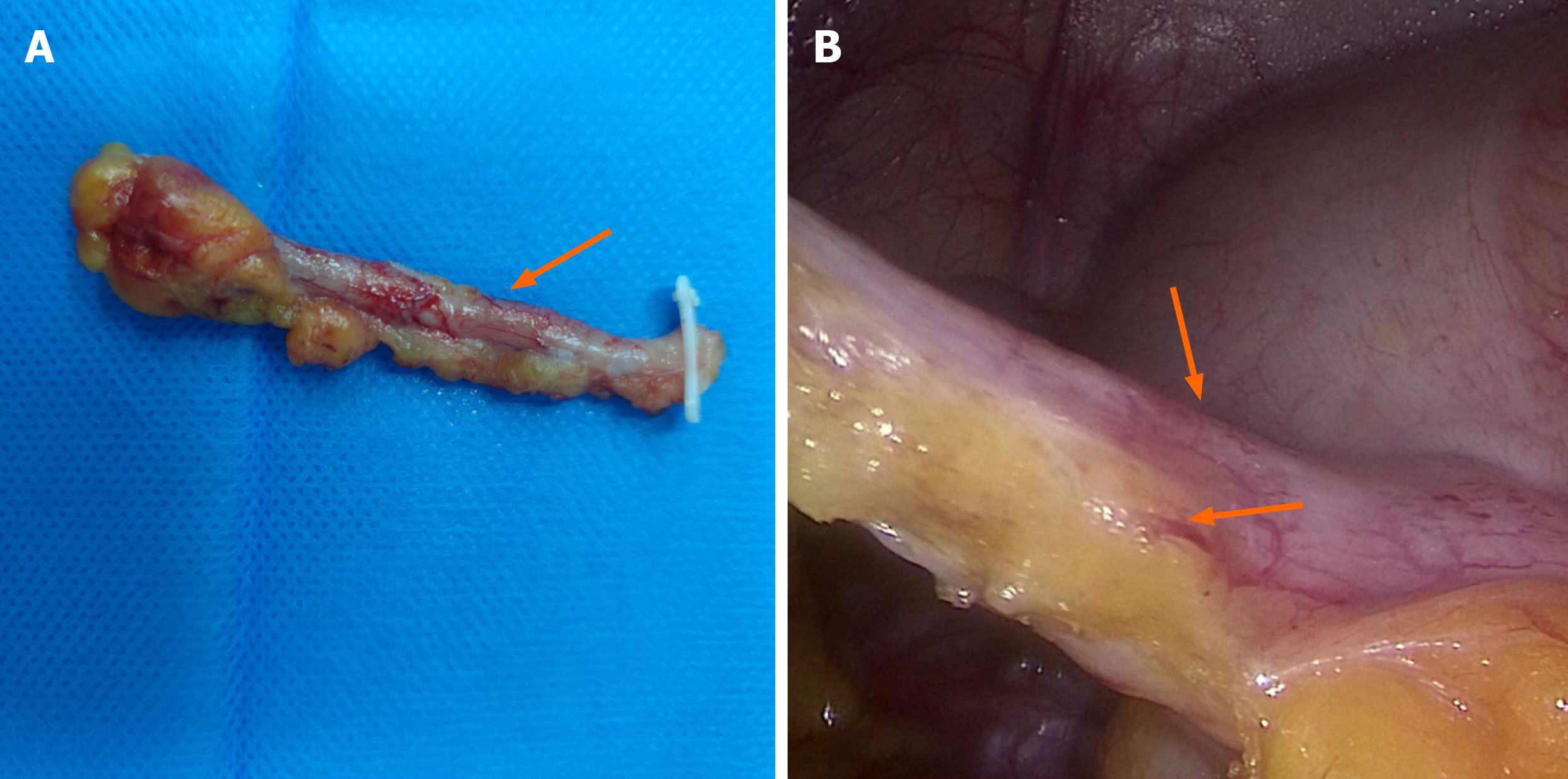
Figure 2 Specimens of the appendix and intraoperative images.
A: The appendix was swollen 3-5 cm from the appendicular root; B: A pulsating blood vessel could be observed in the mesangium of the appendix.
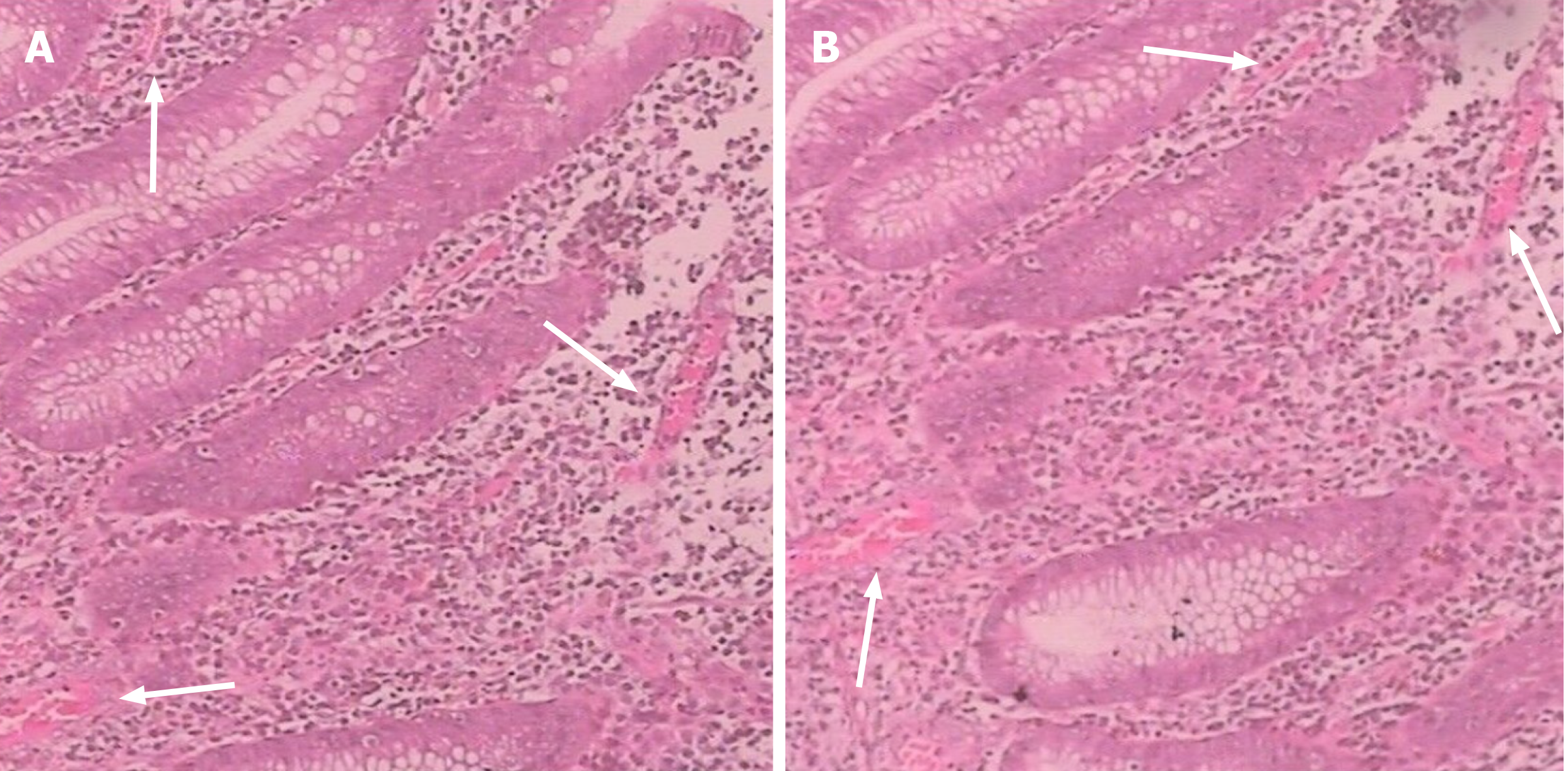
Figure 3 The pathology of the appendix.
A and B: A large number of hyperplastic vessels were observed in the appendix mucosa and capillary vessels were dilated.
- Citation: Ma Q, Du JJ. Appendiceal bleeding caused by vascular malformation: A case report. World J Clin Cases 2024; 12(14): 2457-2462
- URL: https://www.wjgnet.com/2307-8960/full/v12/i14/2457.htm
- DOI: https://dx.doi.org/10.12998/wjcc.v12.i14.2457


