Copyright
©The Author(s) 2024.
World J Clin Cases. May 16, 2024; 12(14): 2426-2430
Published online May 16, 2024. doi: 10.12998/wjcc.v12.i14.2426
Published online May 16, 2024. doi: 10.12998/wjcc.v12.i14.2426
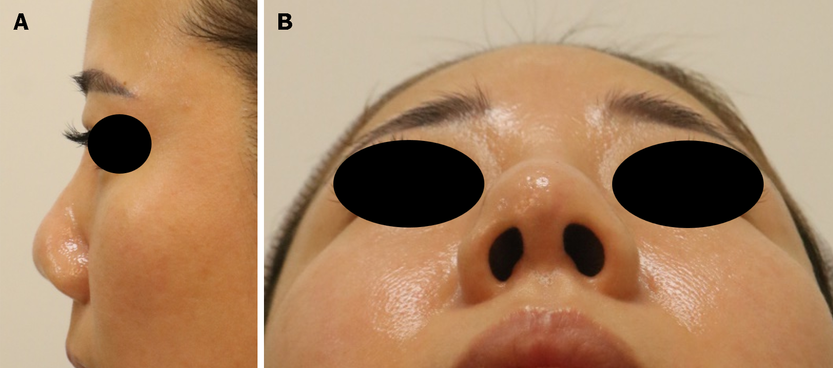
Figure 1 Photograph taken before the secondary rhinoplasty, 40 months after the initial rhinoplasty.
A: Lateral view; B: Worm’s eye view. The skin at the tip of the nose had become thinner, contracture had occurred, and the outline of the implant was visible.
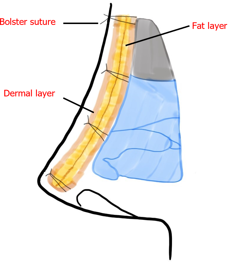
Figure 2 Schematic diagram of the procedure.
The dermofat graft was folded in half, with the fat layer on the inside and the dermal layer facing outward. The folded graft was inserted into the pocket of the nasal dorsum and fixed transcutaneously on the nasal root using a bolster suture.
Figure 3 Photograph taken seven months after the secondary rhinoplasty.
A: Lateral view; B: Worm’s eye view. Despite removing the implant, the nasal height was not significantly depressed and severe deformity did not occur as the contracture at the nasal tip was corrected.
- Citation: Kim H, Kim JH, Koh IC, Lim SY. Immediate secondary rhinoplasty using a folded dermofat graft for resolving complications related to silicone implants: A case report. World J Clin Cases 2024; 12(14): 2426-2430
- URL: https://www.wjgnet.com/2307-8960/full/v12/i14/2426.htm
- DOI: https://dx.doi.org/10.12998/wjcc.v12.i14.2426


