Copyright
©The Author(s) 2024.
World J Clin Cases. May 16, 2024; 12(14): 2420-2425
Published online May 16, 2024. doi: 10.12998/wjcc.v12.i14.2420
Published online May 16, 2024. doi: 10.12998/wjcc.v12.i14.2420
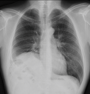
Figure 1 Chest radiograph.
A chest radiograph showed elevation of the right hemidiaphragm.
Figure 2 Computed tomography of the thorax and abdomen.
A: Axial view; B: Coronal view; C: Sagittal view. Computed tomography reveals prolapse of multiple intraabdominal organs into the right thoracic cavity, corresponding to a right-sided Bochdalek hernia. The herniated organs include the stomach, transverse colon, gallbladder, and left lobe of the liver.
Figure 3 Surgical findings.
A and B: The stomach, transverse colon, greater omentum, liver, and gallbladder are herniated into the thoracic cavity via the diaphragmatic defect; C: The left lobe of the liver is completely trapped in the thoracic cavity; D: The diaphragmatic defect is found to be 10 cm × 8 cm; E: The diaphragmatic defect is closed using interrupted sutures; F: The sutured site is reinforced with Bard Composix E/X Mesh®.
- Citation: Mikami S, Kimura S, Tsukamoto Y, Hiwatari M, Hisatsune Y, Fukuoka A, Matsushita T, Enomoto T, Otsubo T. Combined laparoscopic and thoracoscopic repair of adult right-sided Bochdalek hernia with massive liver prolapse: A case report. World J Clin Cases 2024; 12(14): 2420-2425
- URL: https://www.wjgnet.com/2307-8960/full/v12/i14/2420.htm
- DOI: https://dx.doi.org/10.12998/wjcc.v12.i14.2420


