Copyright
©The Author(s) 2024.
World J Clin Cases. May 16, 2024; 12(14): 2404-2411
Published online May 16, 2024. doi: 10.12998/wjcc.v12.i14.2404
Published online May 16, 2024. doi: 10.12998/wjcc.v12.i14.2404
Figure 1 Medical images.
A: Abdominal computed tomography revealed an uneven enhanced lesion located S4/8 segment of liver; B: Abdominal computed tomography revealed a mass in the S6 segment of the liver with a thick calcified wall; C and D: Magnetic resonance images revealed that lesion can be observed at S4/8 segment of liver, with long T1 and mixed with T2 signal.
Figure 2 The procedure of transarterial chemoembolization.
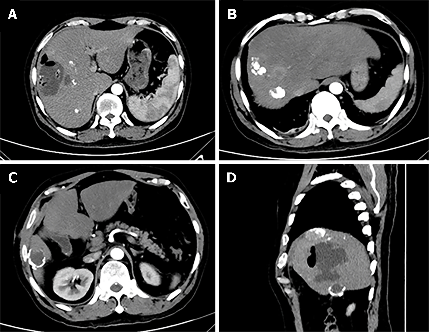
Figure 3 Abdominal computed tomography scanning in the second hospitalization.
A: The plane of liver abscess; B: The plane of hepatocellular carcinoma; C: The plane of hepatic cystic echinococcosis; D: The reconstruction of computed tomography image.
Figure 4 The procedure of percutaneous liver puncture drainage.
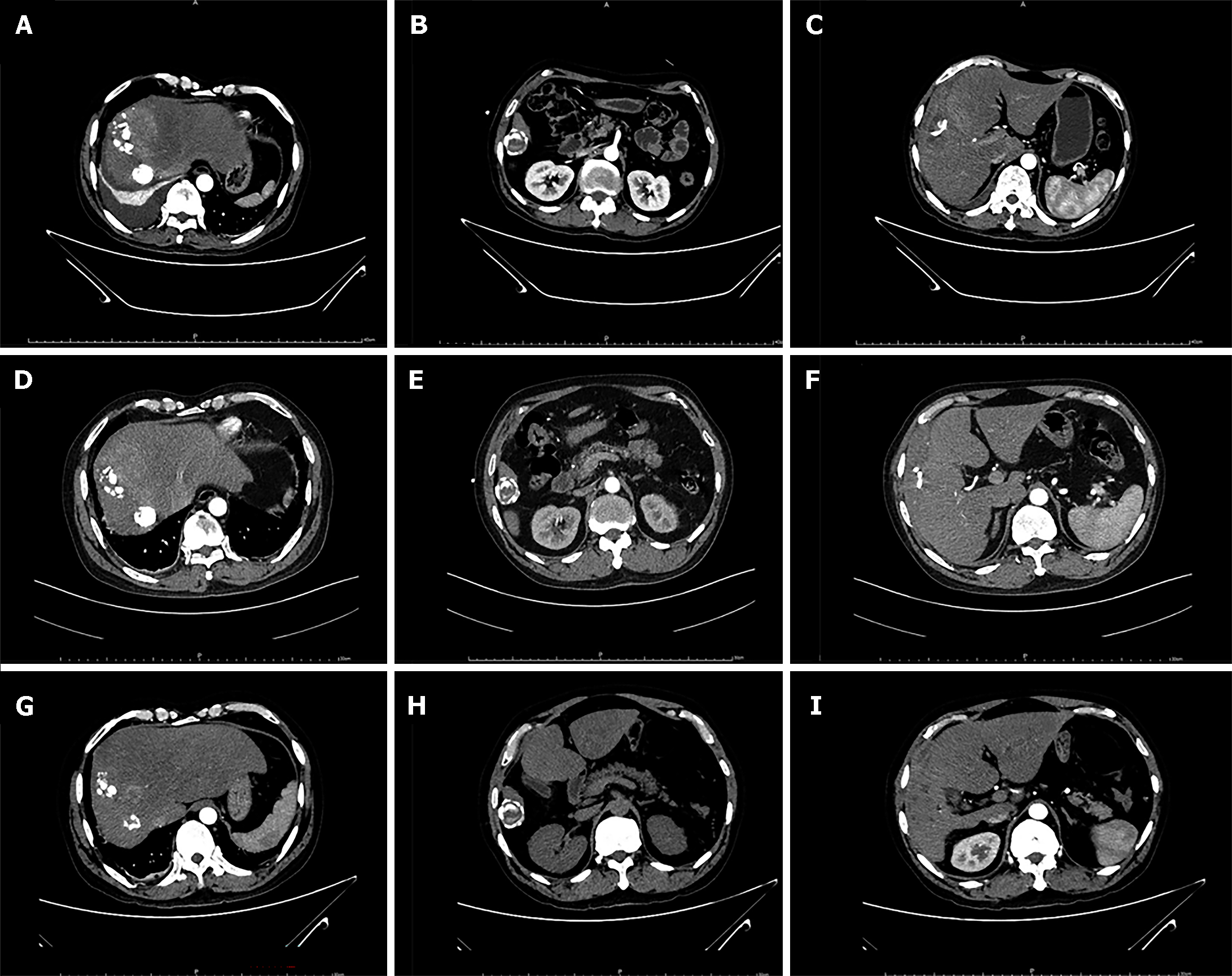
Figure 5 The medical image in the follow-up.
A-C: The plane of hepatocellular carcinoma (HCC), cystic echinococcosis (CE) and liver abscess in June, 2021; D-F: The plane of HCC, CE and liver abscess in October, 2021; G-I: The plane of HCC, CE and liver abscess in April, 2022.
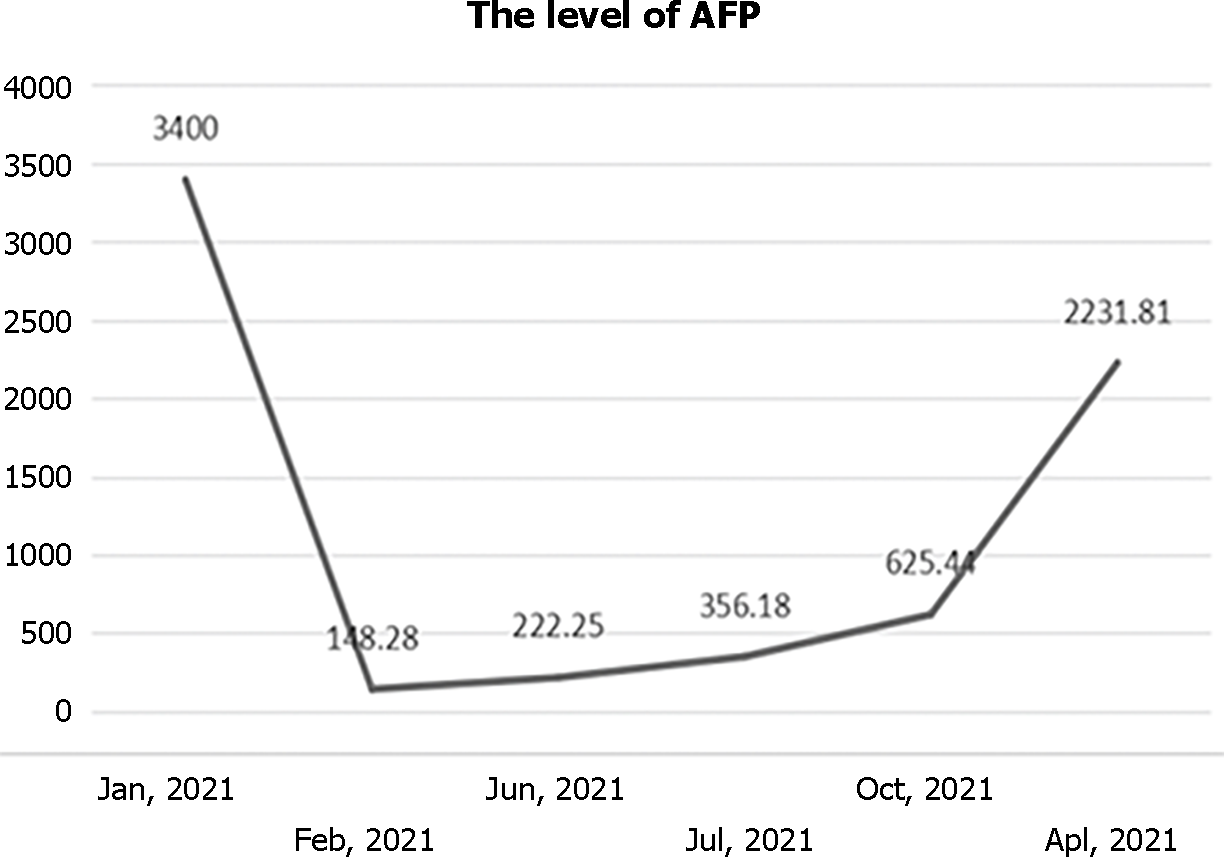
Figure 6 The level of alpha fetoprotein in the follow-up.
AFP: Alpha fetoprotein.
- Citation: Hu YW, Zhao YL, Yan JX, Ma CK. Coexistence of liver abscess, hepatic cystic echinococcosis and hepatocellular carcinoma: A case report. World J Clin Cases 2024; 12(14): 2404-2411
- URL: https://www.wjgnet.com/2307-8960/full/v12/i14/2404.htm
- DOI: https://dx.doi.org/10.12998/wjcc.v12.i14.2404


