Copyright
©The Author(s) 2024.
World J Clin Cases. May 6, 2024; 12(13): 2269-2274
Published online May 6, 2024. doi: 10.12998/wjcc.v12.i13.2269
Published online May 6, 2024. doi: 10.12998/wjcc.v12.i13.2269
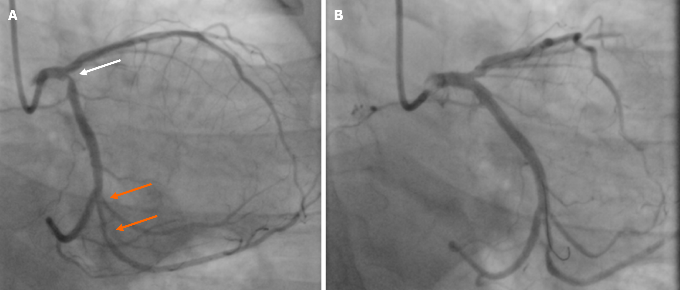
Figure 1 Angiographic images of left coronary artery before (A) and after (B) intervention.
White arrow shows stenosis in the ostial part of circumflex artery stenosis, and two orange arrows depicts 2 tandem stenoses in the obtuse marginal branch. Coronary angiography after stenting from left main to circumflex coronary artery.
Figure 2 Invasive functional parameters of the obtuse marginal stenoses.
A: Coronary stenoses in obtuse marginal branch are shown in orange arrows; B: Corresponding fractional flow reserve, coronary flow reserve, and index of microvascular resistance values.
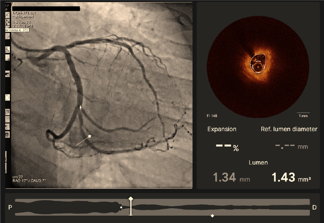
Figure 3 Co-registration images of angiography (left) and optical coherence tomography cross-section imaging (right).
- Citation: Al Nooryani A, Aboushokka W, Beleslin B, Nedeljkovic-Beleslin B. Deferred revascularization in diabetic patient according to combined invasive functional and intravascular imaging data: A case report. World J Clin Cases 2024; 12(13): 2269-2274
- URL: https://www.wjgnet.com/2307-8960/full/v12/i13/2269.htm
- DOI: https://dx.doi.org/10.12998/wjcc.v12.i13.2269


