Copyright
©The Author(s) 2024.
World J Clin Cases. Apr 26, 2024; 12(12): 2128-2133
Published online Apr 26, 2024. doi: 10.12998/wjcc.v12.i12.2128
Published online Apr 26, 2024. doi: 10.12998/wjcc.v12.i12.2128
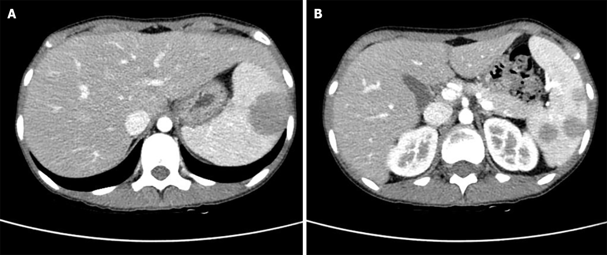
Figure 1 Abdominopelvic computed tomography images of the patient.
A and B: Multiple low-density nodules up to 4 cm in the spleen.
Figure 2 Positron emission tomography-computed tomography images of the patient.
A: Multiple hypermetabolic lymph nodes of various sizes along the left supraclavicular lymph node (SULmax, 3.6); B: Right axillary lymph node (SULmax, 2.7); C: Uneven hypermetabolic activities (SULmax, 3.1) observed in the spleen.
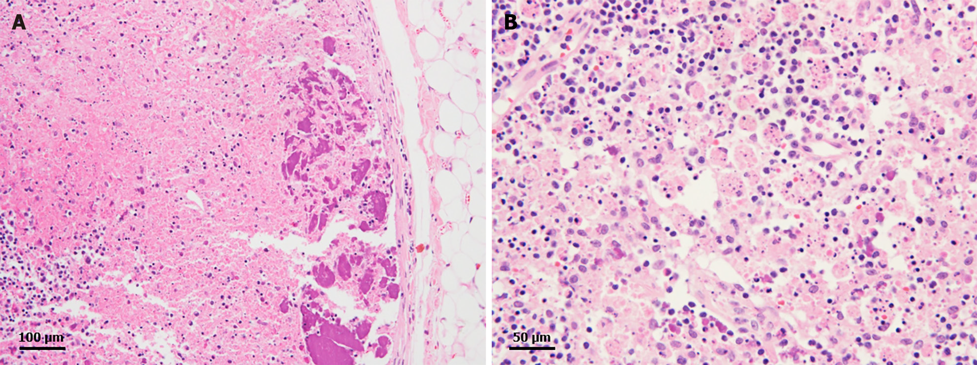
Figure 3 Histopathological findings of the lymph node.
A: Lymph node biopsy reveals extensive subcapsular necrosis with hematoxylin bodies (hematoxylin and eosin stain, × 200); B: Higher magnification shows abundant crescentic histiocytes, abundant karyorrhectic debris, and small hematoxylin bodies (hematoxylin and eosin stain, × 400).
Figure 4 Gross findings after laparoscopic splenectomy.
A and B: Multiple nodules observed on the surface of the spleen. No other specific findings were noted.
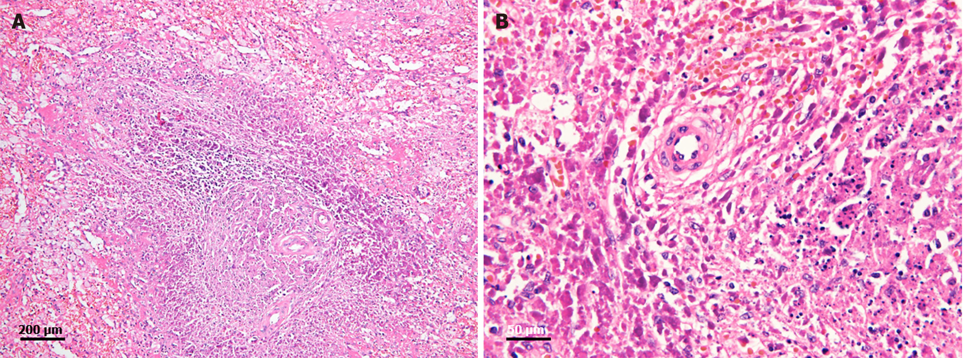
Figure 5 Histological findings of the spleen.
A: Extensive periarteriolar necrosis with hematoxylin bodies (hematoxylin and eosin stain, × 100); B: Higher magnification of hematoxylin bodies and abundant karyorrhectic debris (hematoxylin and eosin stain, × 400).
- Citation: Kang MI, Kwon HC. Systemic lupus erythematosus in a 15-year-old female with multiple splenic nodules: A case report. World J Clin Cases 2024; 12(12): 2128-2133
- URL: https://www.wjgnet.com/2307-8960/full/v12/i12/2128.htm
- DOI: https://dx.doi.org/10.12998/wjcc.v12.i12.2128


