Copyright
©The Author(s) 2024.
World J Clin Cases. Apr 6, 2024; 12(10): 1824-1829
Published online Apr 6, 2024. doi: 10.12998/wjcc.v12.i10.1824
Published online Apr 6, 2024. doi: 10.12998/wjcc.v12.i10.1824
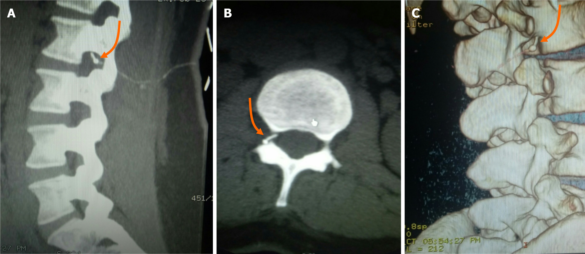
Figure 1 Computed tomography images of the patient.
A-C: Computed tomography images of the lumbar region show a knot in the catheter at the right subvertebral notch of the L2 vertebra (indicated by orange arrows).
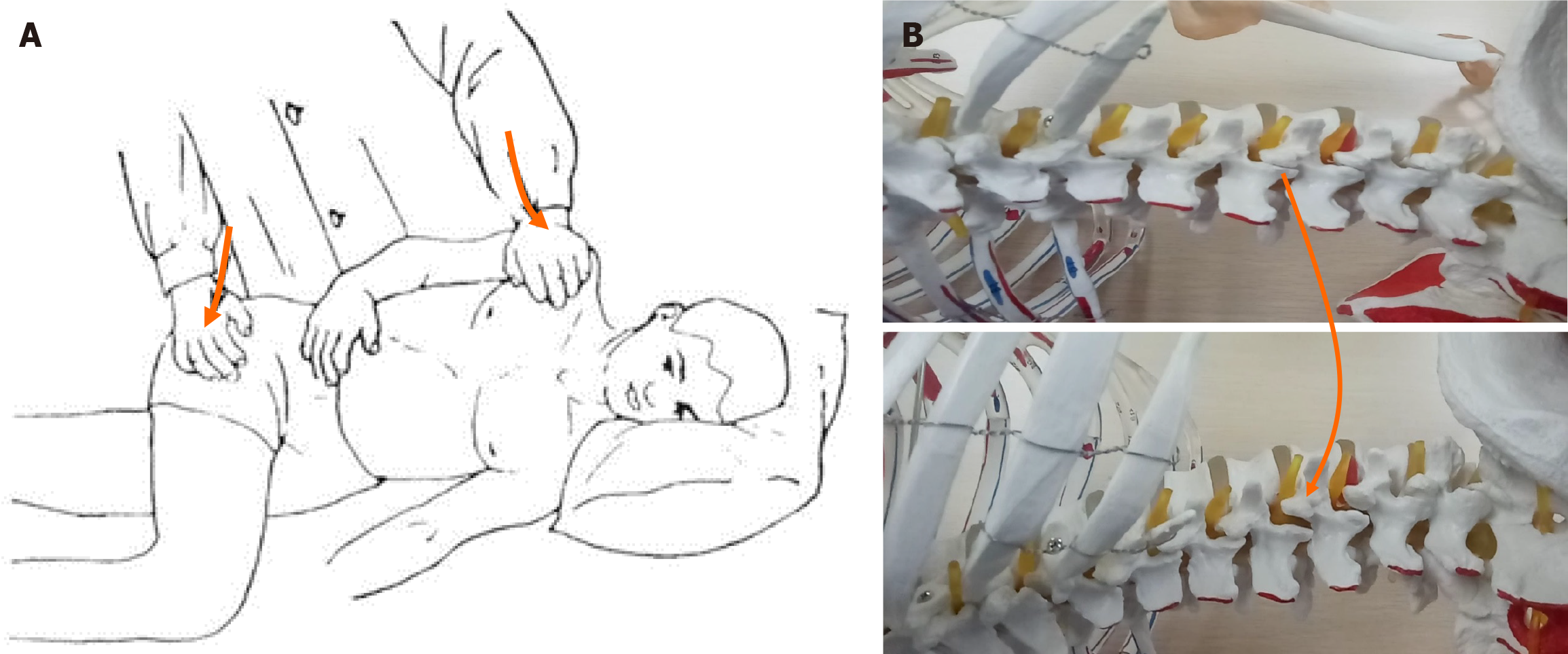
Figure 2 A unique method was adopted in order to pull out the knotted catheter in the patient's epidural space.
A: The patient was placed in a left lateral position with the left lower limb extended and the right lower limb flexed at a 90-degree angle. The operator applied pressure on the patient's right scapula, pushing it backward and downward with the left hand while pushing the patient's right hip joint forward with the right hand; B: Demonstration of a spinal model showing the steps presented in A, which effectively opened the right-sided facet joint of the lumbar vertebrae.
Figure 3 The shape of the knotted catheter in the epidural space after it was successfully pulled out.
A knot located approximately 3.2 cm from the catheter tip (indicated by the orange arrow). The soft portion of the catheter coil fractured at a distance of 8 cm from the catheter tip (indicated by the yellow arrow).
- Citation: Deng NH, Chen XC, Quan SB. Unique method for removal of knotted lumbar epidural catheter: A case report. World J Clin Cases 2024; 12(10): 1824-1829
- URL: https://www.wjgnet.com/2307-8960/full/v12/i10/1824.htm
- DOI: https://dx.doi.org/10.12998/wjcc.v12.i10.1824


