Copyright
©The Author(s) 2024.
World J Clin Cases. Apr 6, 2024; 12(10): 1772-1777
Published online Apr 6, 2024. doi: 10.12998/wjcc.v12.i10.1772
Published online Apr 6, 2024. doi: 10.12998/wjcc.v12.i10.1772
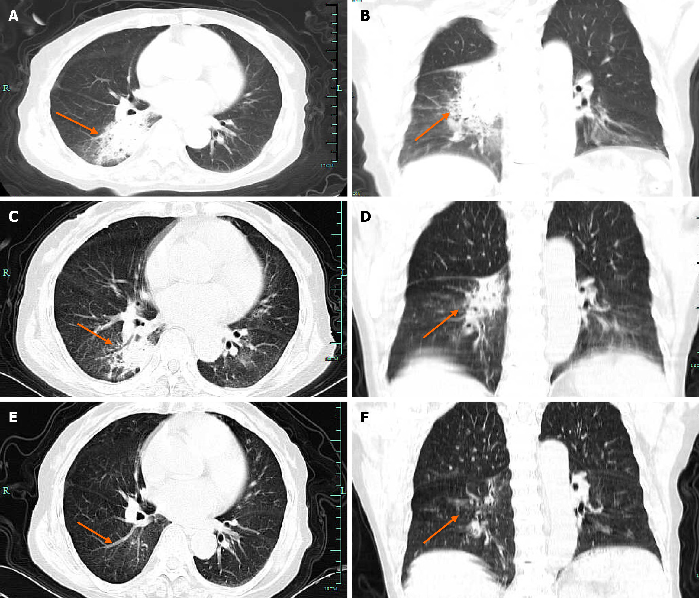
Figure 1 Chest computed tomography on admission and post-treatment.
A: Pulmonary consolidation in the posterior segment of the right lower lobe at admission [axial thorax computed tomography (CT) view); B: Mass consolidation in the posterior segment of the right lower lobe at admission (coronal thorax CT view); C: Lesion reduction in pulmonary consolidation in the posterior segment of the right lower lobe after 2 wk of isavuconazole treatment (axial thorax CT view); D: Lesion reduction in pulmonary consolidation in the posterior segment of the right lower lobe after 2 wk of isavuconazole treatment (coronal thorax CT view); E: Significant absorption of pulmonary consolidation in the posterior segment of the right lower lobe at 6 wk of discharge (axial thorax CT view); F: Significant absorption of pulmonary consolidation in the posterior segment of the right lower lobe at 6 wk of discharge (coronal thorax CT view).
Figure 2 Culture and microscopic examination of the microorganisms.
A: Salmonella glucose agar, 28 °C, culture for 7 d, the purplish colony were concentric on the front; B: Yellowish-pale colonies on the back; C: Colorless hyaline branching filaments were observed under light microscopy, and the spores were erect, with a broomy tip and a cone-shaped, fine bony tip at the base of the flask. The spores were mainly oval or near-spherical in shape, and the chains were cylindrical or discrete.
- Citation: Yang XL, Zhang JY, Ren JM. Successful treatment of Purpureocillium lilacinum pulmonary infection with isavuconazole: A case report. World J Clin Cases 2024; 12(10): 1772-1777
- URL: https://www.wjgnet.com/2307-8960/full/v12/i10/1772.htm
- DOI: https://dx.doi.org/10.12998/wjcc.v12.i10.1772


