Copyright
©The Author(s) 2024.
World J Clin Cases. Jan 6, 2024; 12(1): 188-195
Published online Jan 6, 2024. doi: 10.12998/wjcc.v12.i1.188
Published online Jan 6, 2024. doi: 10.12998/wjcc.v12.i1.188
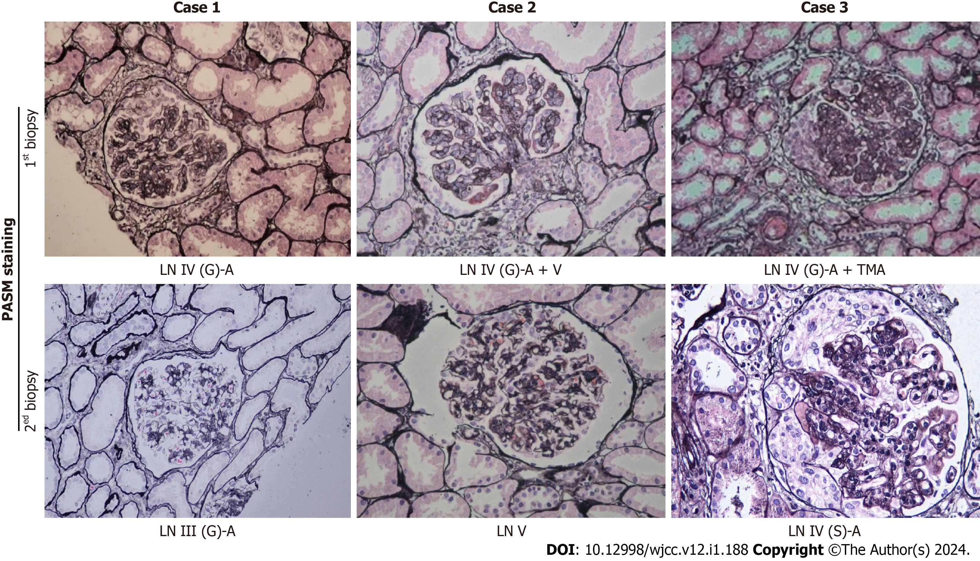
Figure 1 Pathological images of renal biopsy from the three patients (periodic acid-silver methenamine staining, 200 × magnification).
PASM: Periodic acid-silver methenamine; LN: Lupus nephritis; TMA: Thrombotic microangiopathy; G: Global; A: Active; S: Segmental.
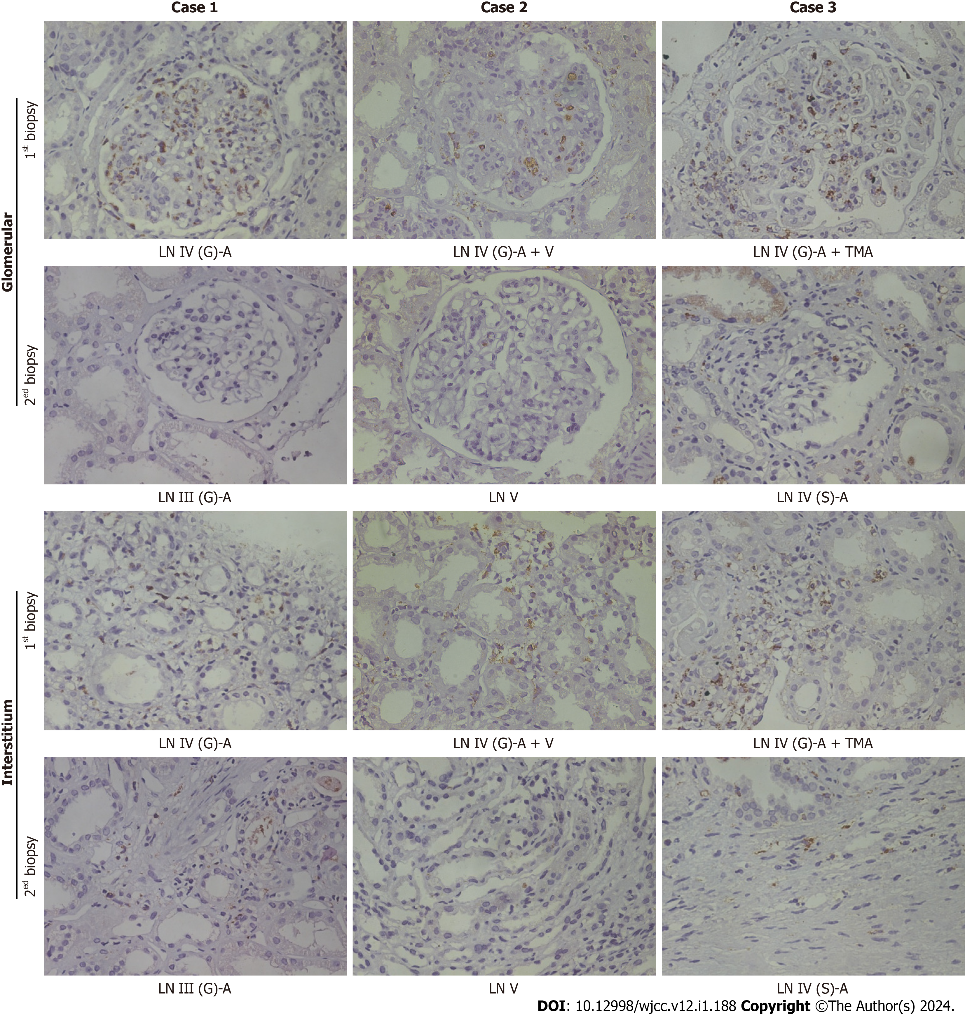
Figure 2 Immunohistochemical staining for CD68-positive macrophages in the glomerulus and renal interstitium.
LN: Lupus nephritis; TMA: Thrombotic microangiopathy; G: Global; A: Active; S: Segmental.
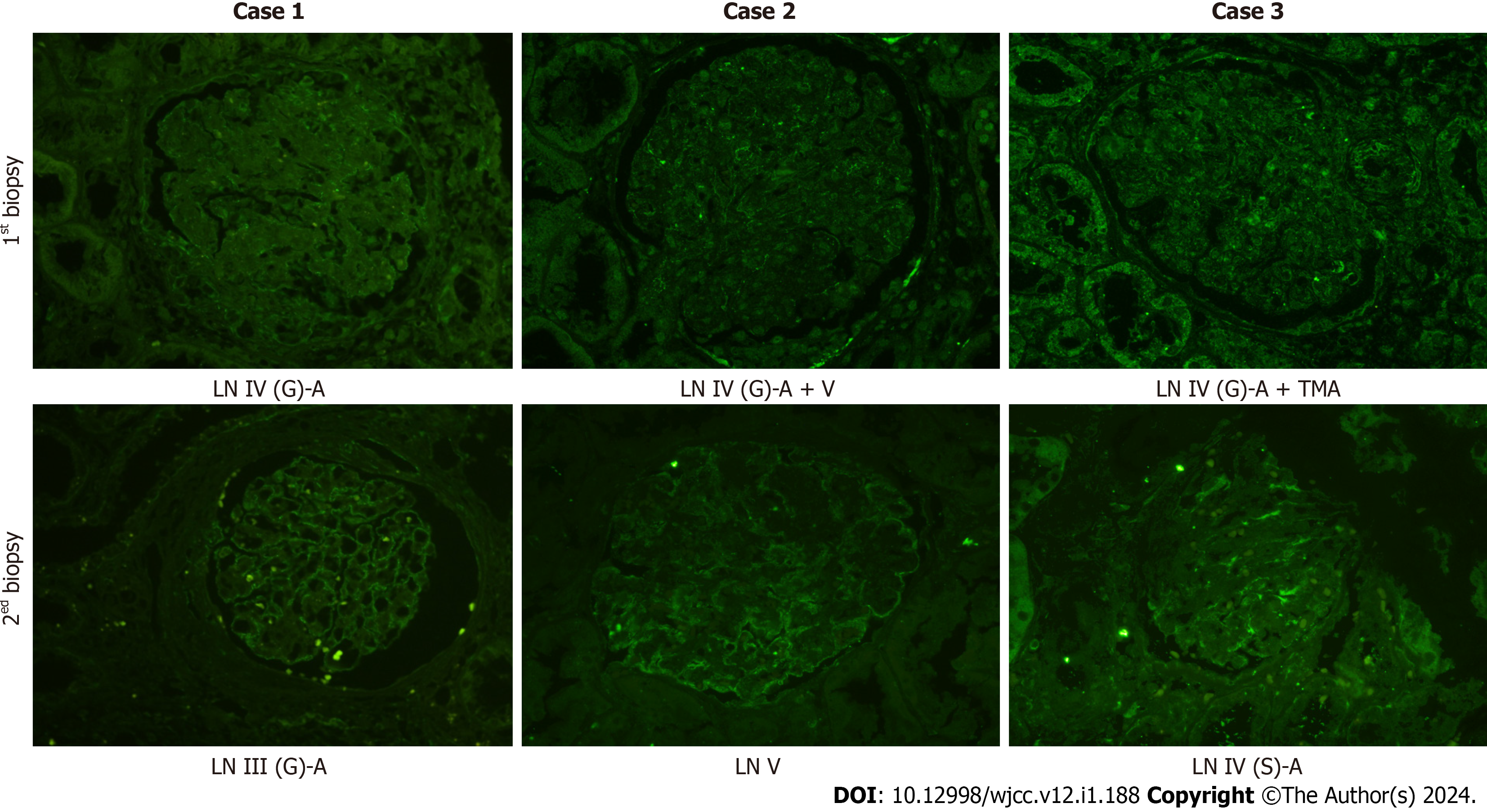
Figure 3 Podocin expression in the glomerulus.
LN: Lupus nephritis; TMA: Thrombotic microangiopathy; G: Global; A: Active; S: Segmental.
- Citation: Liu SY, Chen H, He LJ, Huang CK, Wang P, Rui ZR, Wu J, Yuan Y, Zhang Y, Wang WJ, Wang XD. Changes in macrophage infiltration and podocyte injury in lupus nephritis patients with repeated renal biopsy: Report of three cases. World J Clin Cases 2024; 12(1): 188-195
- URL: https://www.wjgnet.com/2307-8960/full/v12/i1/188.htm
- DOI: https://dx.doi.org/10.12998/wjcc.v12.i1.188


