Copyright
©The Author(s) 2024.
World J Clin Cases. Jan 6, 2024; 12(1): 180-187
Published online Jan 6, 2024. doi: 10.12998/wjcc.v12.i1.180
Published online Jan 6, 2024. doi: 10.12998/wjcc.v12.i1.180
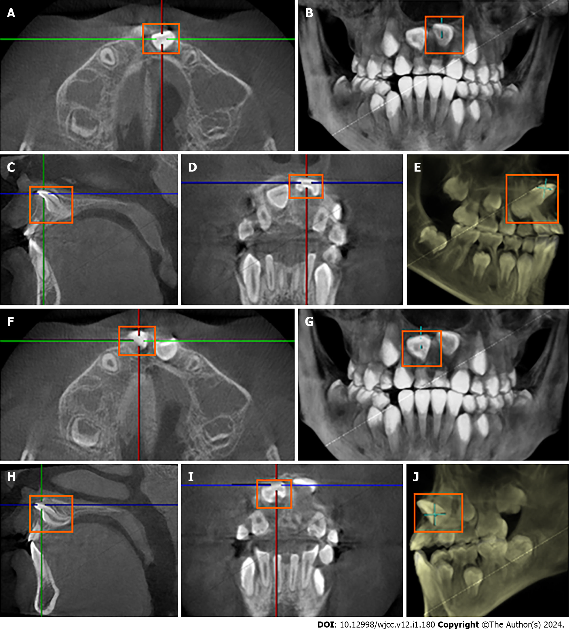
Figure 1 Cone-beam computed tomography images before treatment showing dilaceration of maxillary central incisors.
A and F: Axial cone-beam computed tomography (CBCT) images; B and G: 3D reconstruction-axial plane; C and H: Sagittal CBCT images; D and I: Labial CBCT images; E and J: 3D reconstruction-sagittal plane.
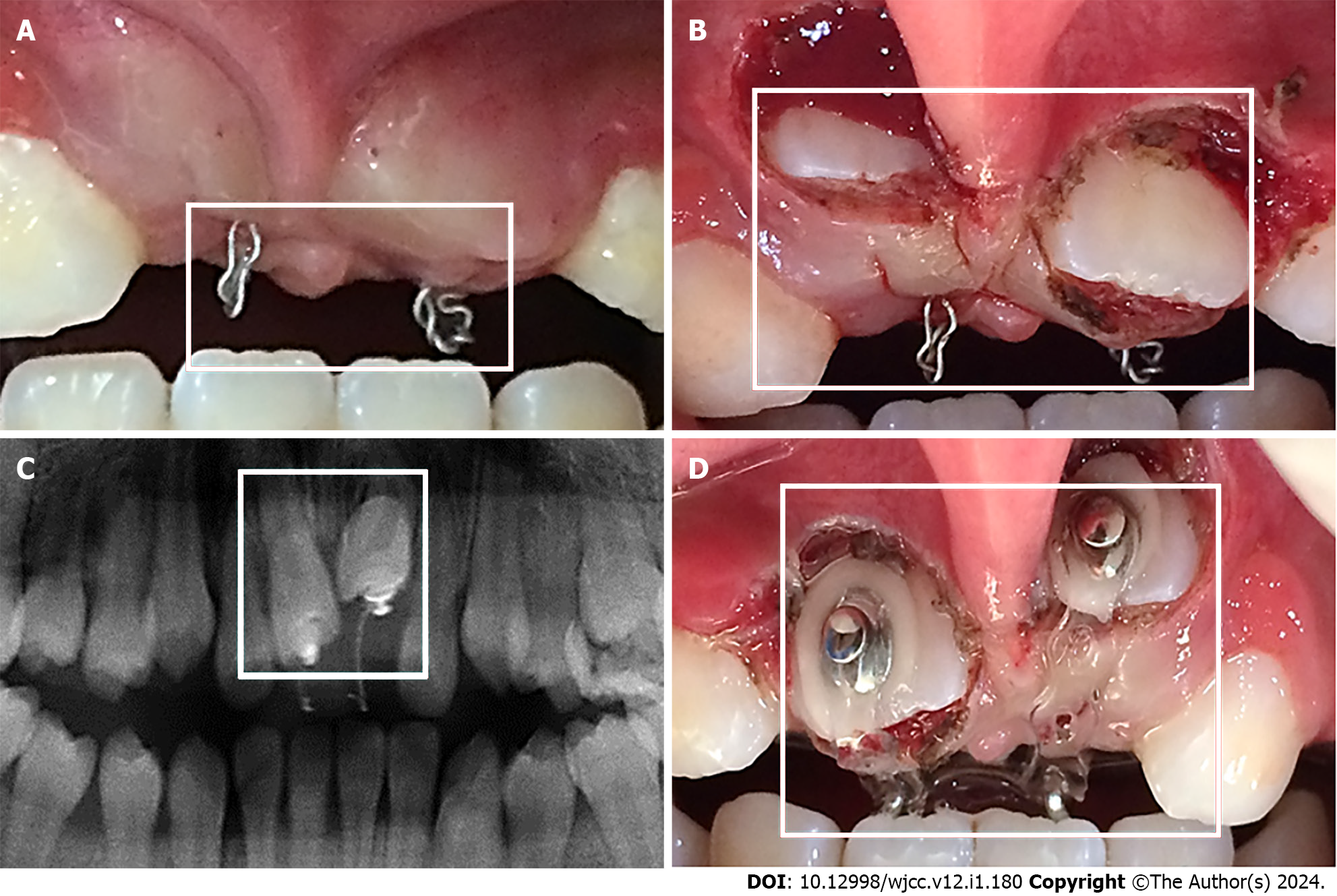
Figure 2 Surgical procedure and tooth traction.
A: Photograph of closed-eruption traction; B: Photograph of the buccal surface exposed by surgery; C: X-ray of closed eruption traction; D: Photograph of the exposed crown with a lingual button bonded to the labial surface.
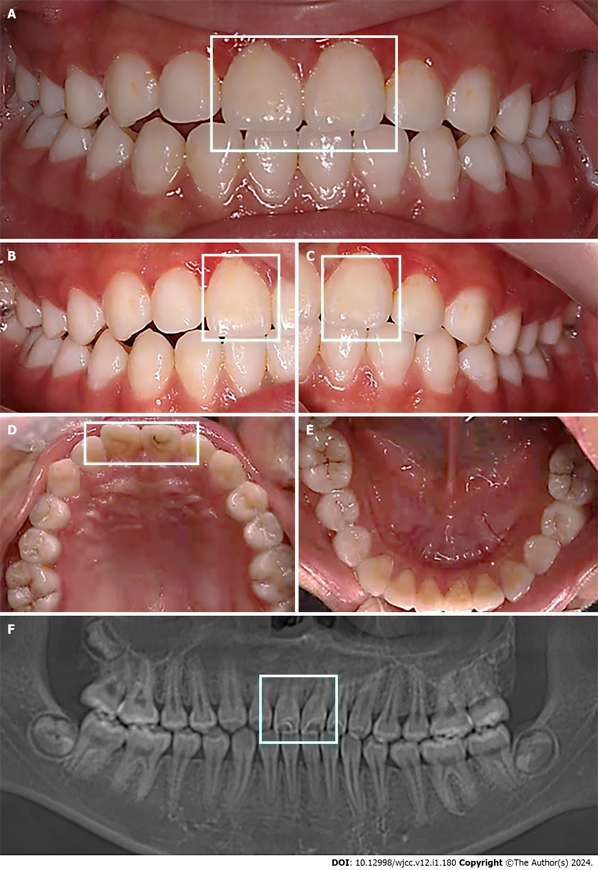
Figure 3 Images after debonding.
A-E: Intraoral photographs; F: Posttreatment panoramic radiograph.
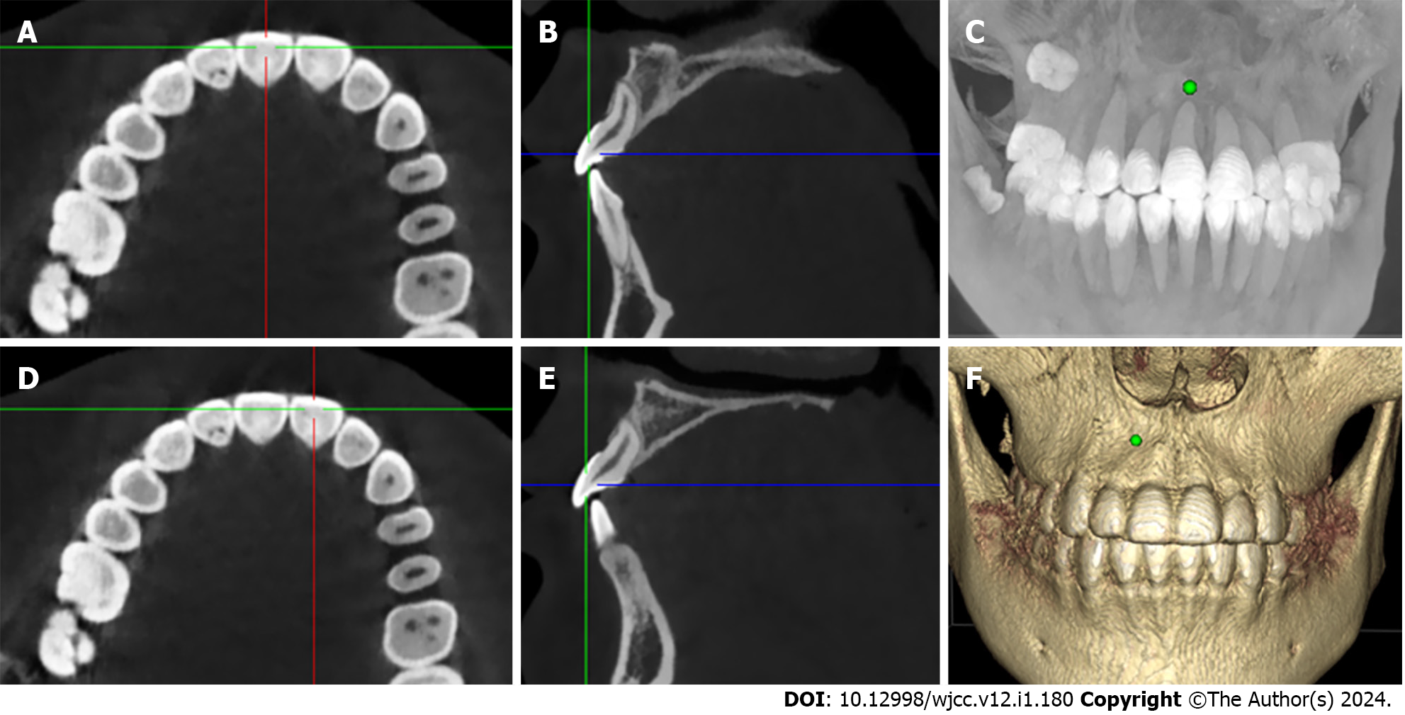
Figure 4 Cone-beam computed tomography images at 3-year follow-up.
A and D: Axial cone-beam computed tomography (CBCT) images; B and E: Sagittal CBCT images; C and F: 3D reconstruction.
- Citation: Wang JM, Guo LF, Ma LQ, Zhang J. Labial inverse dilaceration of bilateral maxillary central incisors: A case report. World J Clin Cases 2024; 12(1): 180-187
- URL: https://www.wjgnet.com/2307-8960/full/v12/i1/180.htm
- DOI: https://dx.doi.org/10.12998/wjcc.v12.i1.180


