Copyright
©The Author(s) 2024.
World J Clin Cases. Jan 6, 2024; 12(1): 163-168
Published online Jan 6, 2024. doi: 10.12998/wjcc.v12.i1.163
Published online Jan 6, 2024. doi: 10.12998/wjcc.v12.i1.163
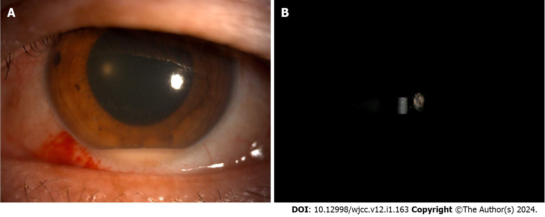
Figure 1 Silt-lamp examination on the second day after surgery.
A: Hypopyon in the anterior chamber; B: Cells in the anterior chamber.
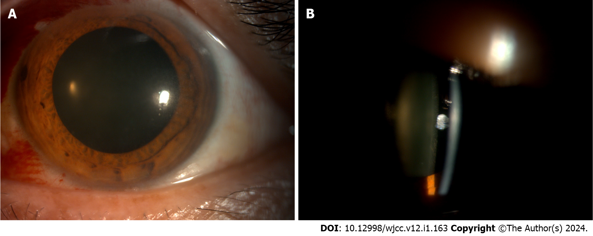
Figure 2 Silt-lamp examination on the fifth day after surgery.
A: Decreased hypopyon; B: Fold of corneal posterior elastic layer with decreased hypopyon.
Figure 3 Silt-lamp examination one week postoperatively.
A: A clear anterior chamber in diffuse light; B: A clear anterior chamber in silt light.
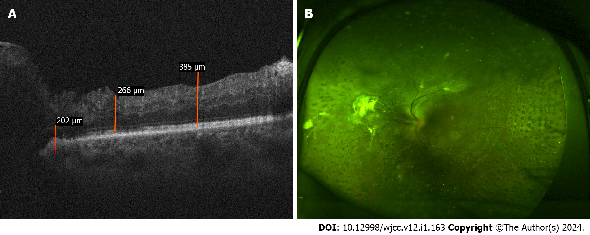
Figure 4 Optical coherence tomography and fundus examination 3 mo postoperatively.
A: Diabetic macular edema in the nasal region, with most of the structure being normal; B: Reattached retina.
- Citation: Yan HC, Wang ZL, Yu WZ, Zhao MW, Liang JH, Yin H, Shi X, Miao H. Endophthalmitis in silicone oil-filled eye: A case report. World J Clin Cases 2024; 12(1): 163-168
- URL: https://www.wjgnet.com/2307-8960/full/v12/i1/163.htm
- DOI: https://dx.doi.org/10.12998/wjcc.v12.i1.163


