Copyright
©The Author(s) 2024.
World J Clin Cases. Jan 6, 2024; 12(1): 148-156
Published online Jan 6, 2024. doi: 10.12998/wjcc.v12.i1.148
Published online Jan 6, 2024. doi: 10.12998/wjcc.v12.i1.148
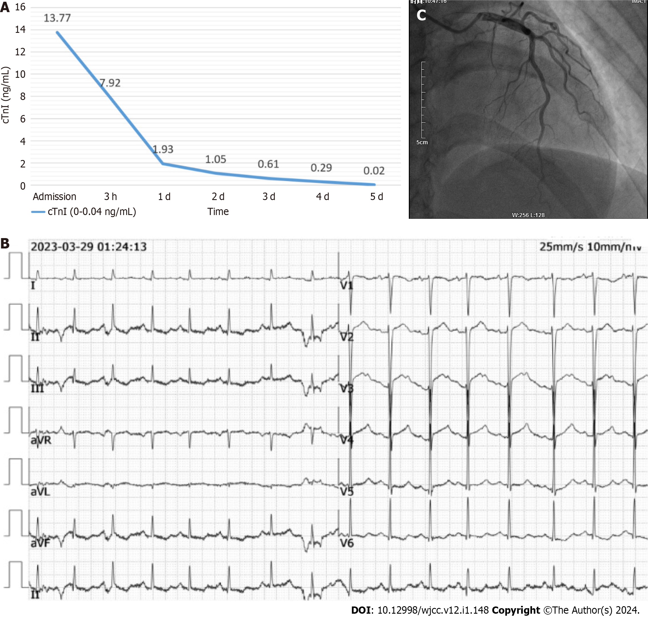
Figure 1 Patient examination data on admission.
A: The patient's levels of myocardial troponin were elevated upon admission, reaching over 300 times the reference upper limit; B: Electrocardiogram shows an ST-segment elevation of 2 mm in leads V2, V3, and V4; C: Coronary angiography did not reveal any signs of coronary artery spasms.
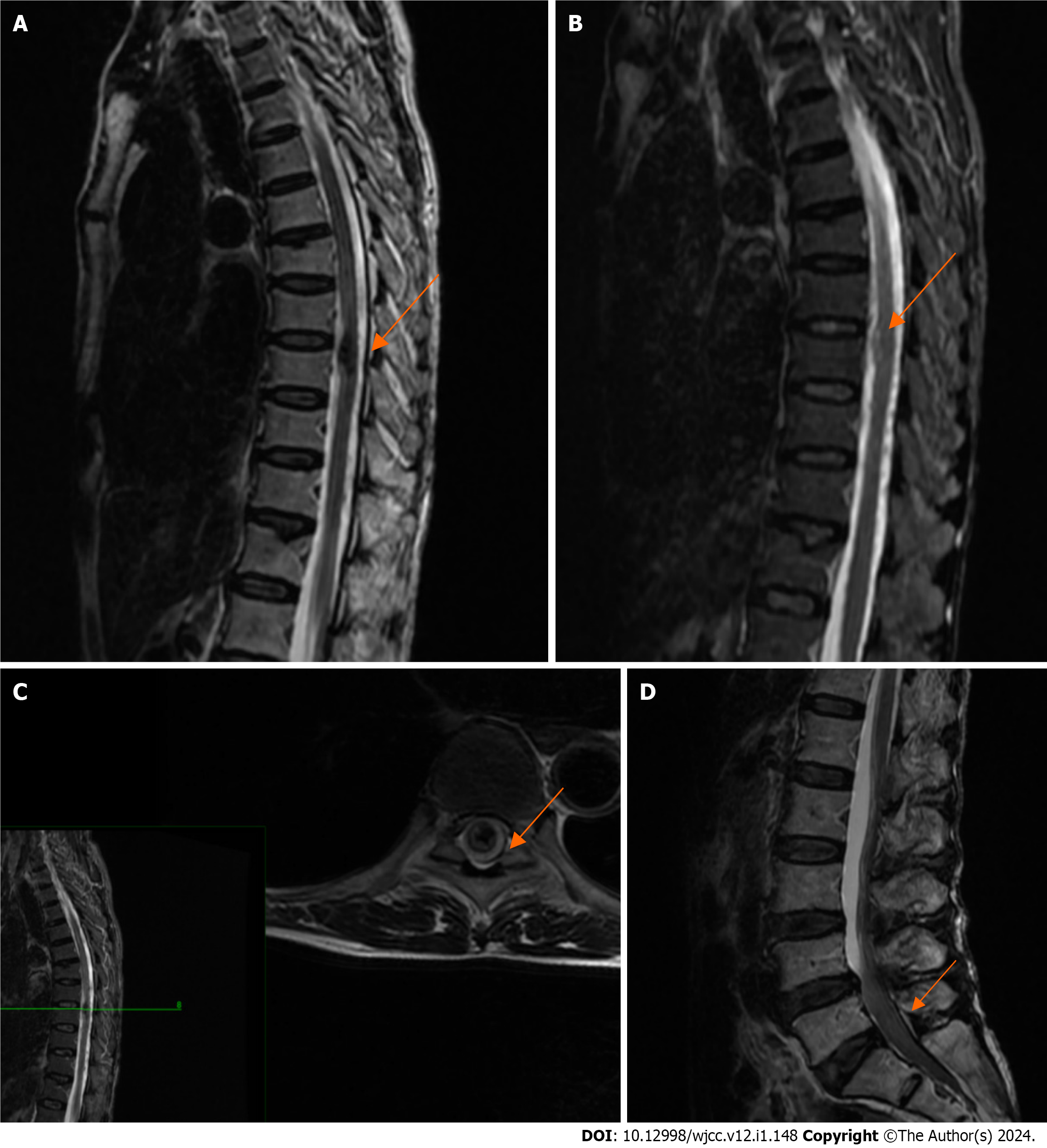
Figure 2 Preoperative magnetic resonance imaging of the thoracic and lumbar spin.
A-C: Magnetic resonance imaging (MRI) of the thoracic spine was considered for acute bleeding in the T9 segment of the spinal cord; D: MRI of the lumbar spine indicated abnormal signals in the spinal canal at the L4-S2 segment, suggesting the presence of a hematoma.
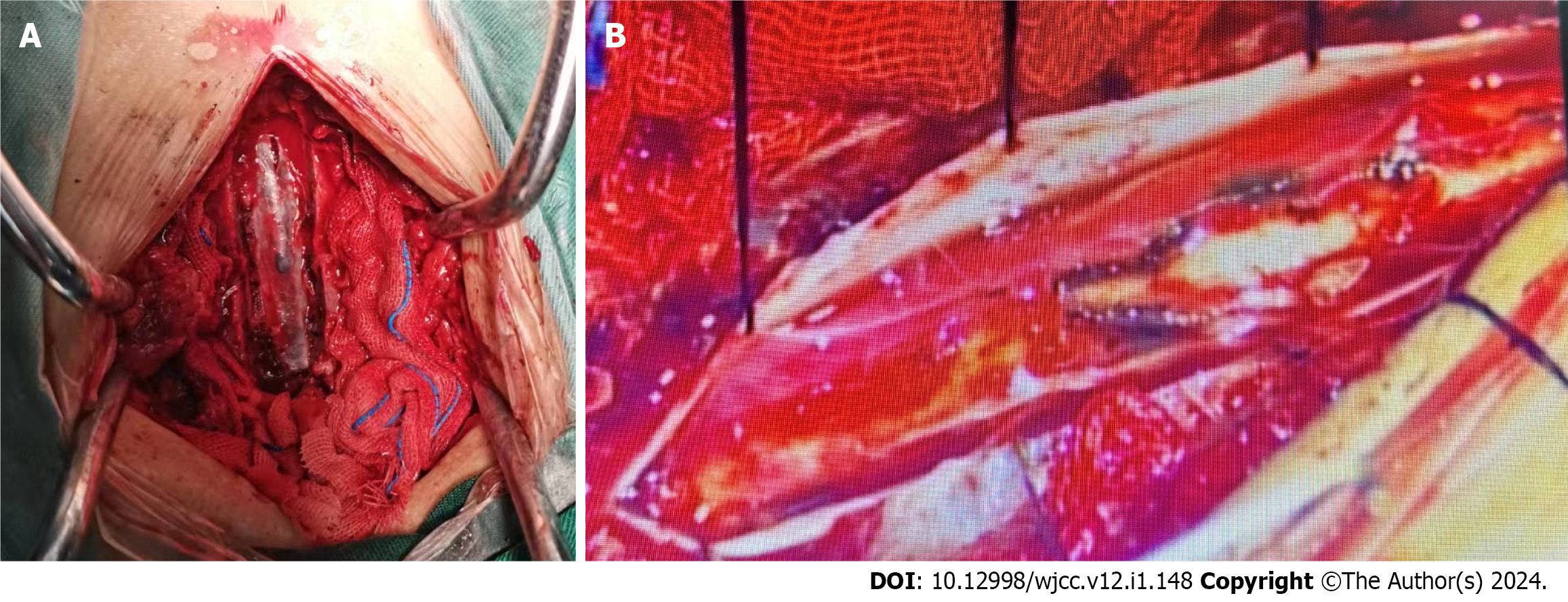
Figure 3 Intravertebral canal of the thoracic spine seen intraoperatively.
A: Intraoperatively, diffuse hematoma formation was found in the T7-T10 spinal canal, and no significant spinal vascular malformation changes were found; B: Under the microscope, the arachnoid membrane of the spinal cord was detached, the hematoma was completely removed, and bleeding was stopped thoroughly.
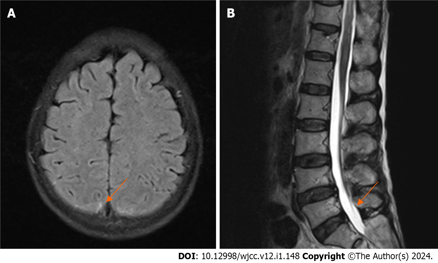
Figure 4 Postoperative magnetic resonance imaging of the lumbar spine and brain.
A: Cranial magnetic resonance imaging (MRI) showed a small amount of subarachnoid hemorrhage; B: Postoperative lumbar MRI showed a redistribution of the intraspinal hematoma.
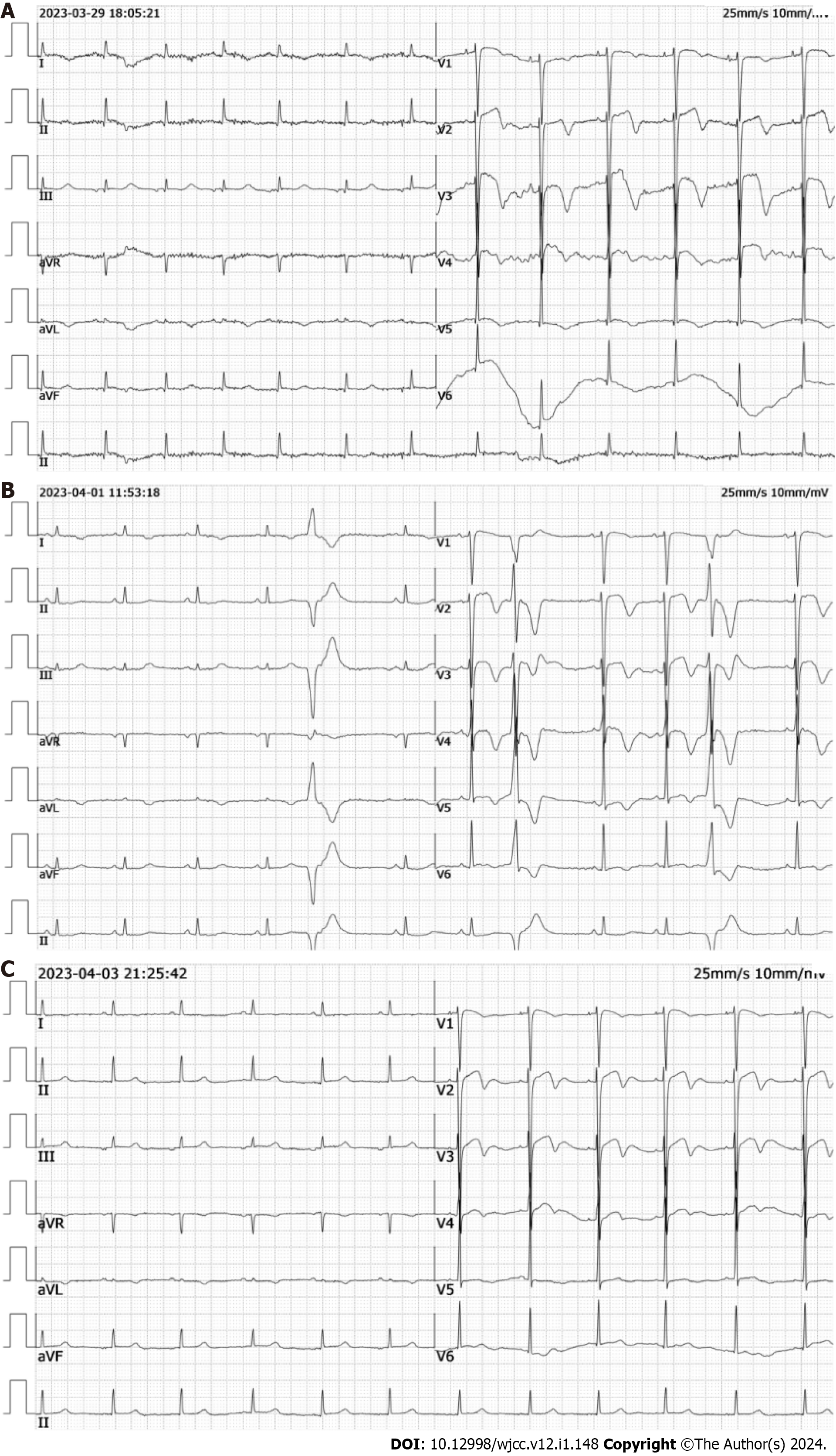
Figure 5 Electrocardiograms.
A: ST-segment elevation and T-wave inversion in leads V2, V3, and V4; B: ST-segment elevation and T-wave inversion are more pronounced, with no pathologic Q waves; C: ST-segment elevation and T-wave inversion have decreased compared to previous readings.
- Citation: Lin JM, Yuan XJ, Li G, Gan XR, Xu WH. Subarachnoid hemorrhage misdiagnosed as acute coronary syndrome leading to catastrophic neurologic injury: A case report. World J Clin Cases 2024; 12(1): 148-156
- URL: https://www.wjgnet.com/2307-8960/full/v12/i1/148.htm
- DOI: https://dx.doi.org/10.12998/wjcc.v12.i1.148


