Copyright
©The Author(s) 2023.
World J Clin Cases. Mar 26, 2023; 11(9): 2110-2115
Published online Mar 26, 2023. doi: 10.12998/wjcc.v11.i9.2110
Published online Mar 26, 2023. doi: 10.12998/wjcc.v11.i9.2110
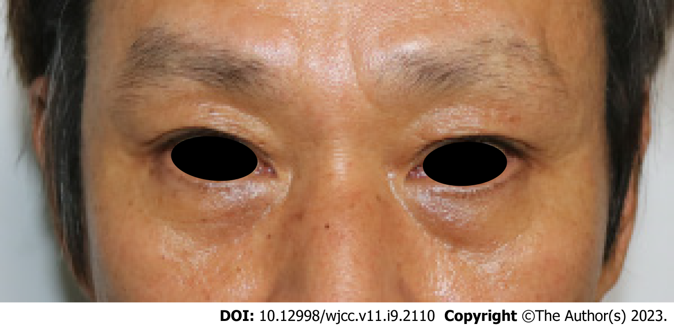
Figure 1 A facial photograph taken at the moment of admission.
The swelling around the periorbital area was not significant.
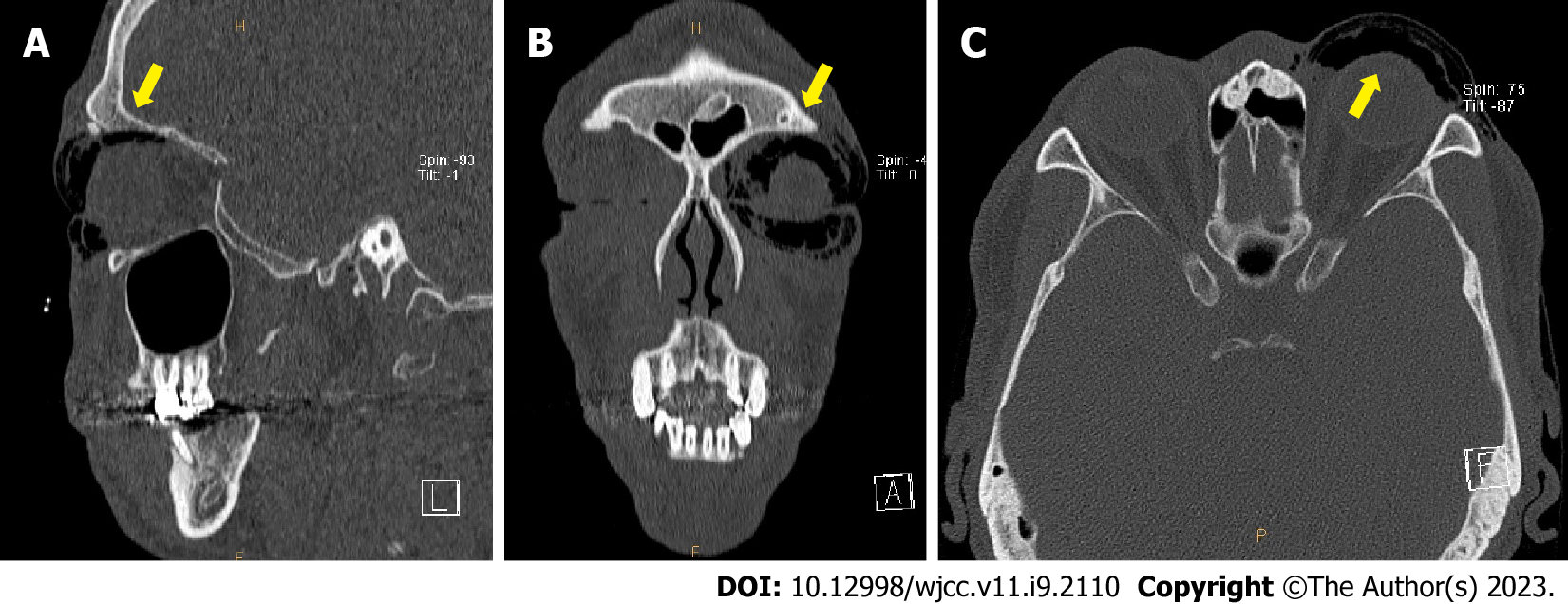
Figure 2 A Follow up computed tomography scan 2 d postoperatively.
The emphysema is seen in the form of scattered air bubbles in the left periorbital subcutaneous area (yellow arrow), indicating the “black eyebrow sign’. A: Sagittal view; B: Coronal view; C: Axial view.
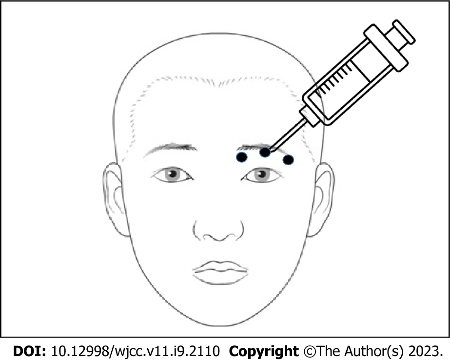
Figure 3 A schematic drawing of needle injection point (black circles).
Above the upper eyelid, the needle was inserted at points where air was most palpable and negative pressure was applied using the pull-back method.
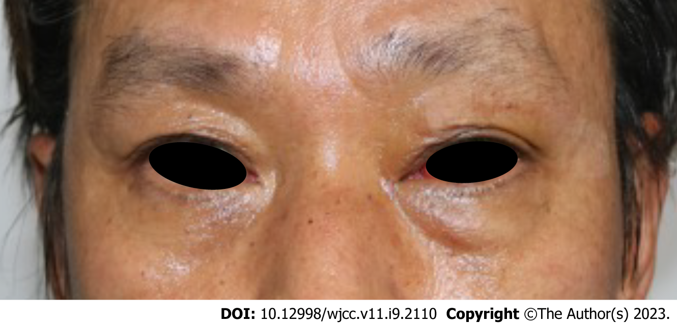
Figure 4 A facial photograph taken after the procedure.
A: One day postoperatively, swelling in the left periorbital area was observed; B: The swelling improved immediately after needle aspiration decompression.
Figure 5 A facial photograph 4 d postoperatively.
No recurrence of swelling was observed.
- Citation: Nam HJ, Wee SY. Successful treatment of a rare subcutaneous emphysema after a blow-out fracture surgery using needle aspiration: A case report. World J Clin Cases 2023; 11(9): 2110-2115
- URL: https://www.wjgnet.com/2307-8960/full/v11/i9/2110.htm
- DOI: https://dx.doi.org/10.12998/wjcc.v11.i9.2110


