Copyright
©The Author(s) 2023.
World J Clin Cases. Mar 26, 2023; 11(9): 2104-2109
Published online Mar 26, 2023. doi: 10.12998/wjcc.v11.i9.2104
Published online Mar 26, 2023. doi: 10.12998/wjcc.v11.i9.2104
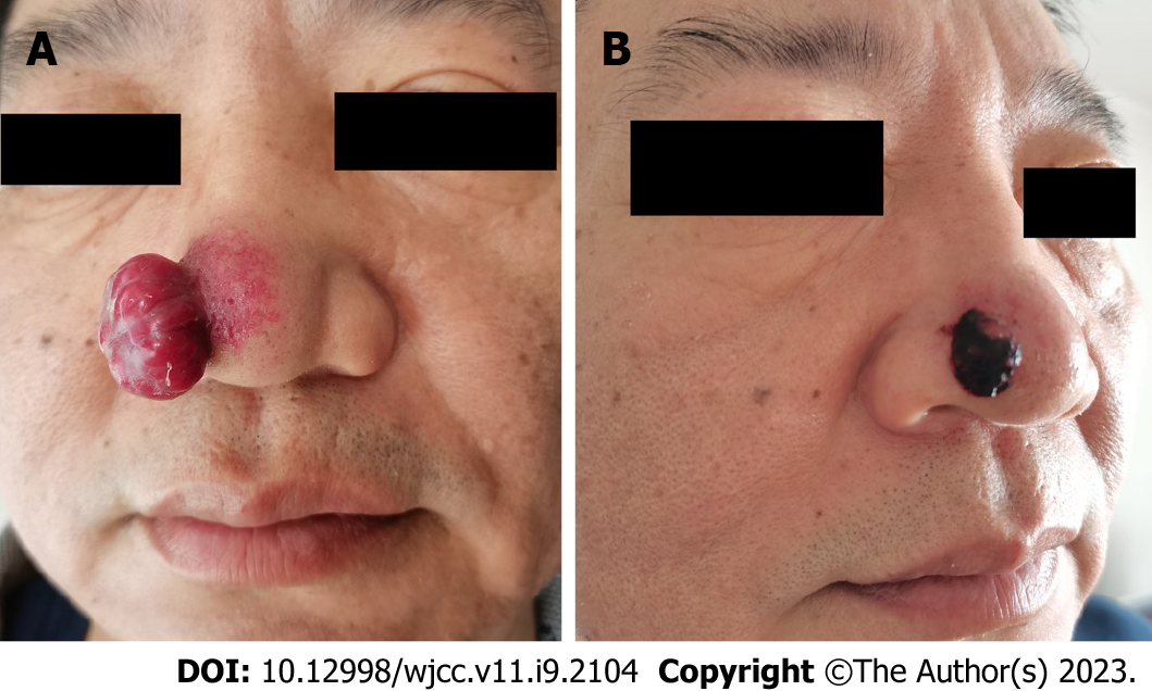
Figure 1 Clinical photograph of the lesion at the initial visit and after resumption of target therapy.
A: A 2.0 cm × 2.0 cm × 1.2 cm red mass was seen on the right alar of the nose, with soft texture and clear boundary. A small amount of bloody exudate was seen on the surface. The skin vessels around the mass were hyperplastic and dilated; B: Two weeks after the resumption of combined treatment, the mass was reduced to 0.8 cm × 0.8 cm × 0.5 cm, covered with blood scabs, and the hyperplasia and dilation of skin vessels around the mass decreased.
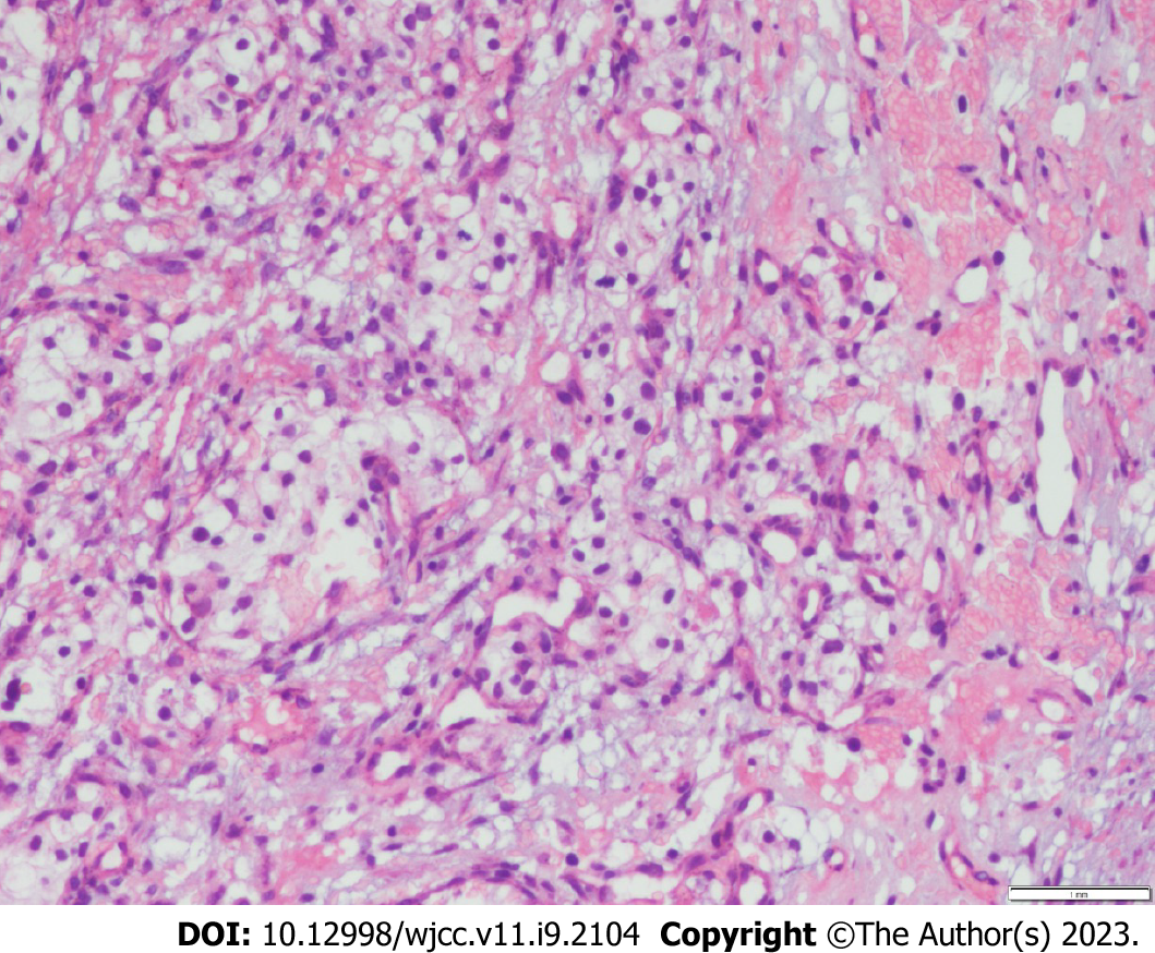
Figure 2 The histopathology of the mass (hematoxylin & eosin staining).
Lumplike infiltration of tumor cells was observed in the dermis, some of which formed lumenlike structures with a large number of clear cells and abundant blood vessels.
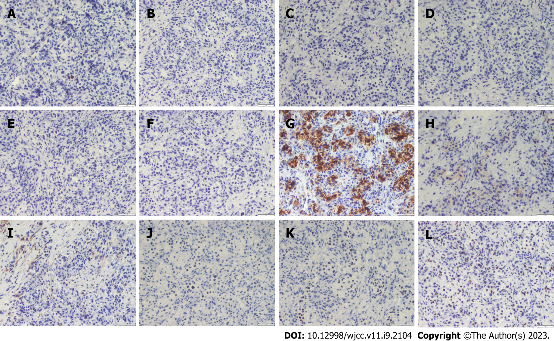
Figure 3 Immunopathology of the mass.
A: S100(-); B: HMB45(-); C: CK7(-); D: AR(-); E: CK5/6(-); F: CAIX(-); G: Vimentin(+); H: EMA(+); I: CD10(+); J: PAX8(+); K: PAX2(+); L: Ki67(+).
- Citation: Dong S, Xu YC, Zhang YC, Xia JX, Mou Y. Pembrolizumab combined with axitinib in the treatment of skin metastasis of renal clear cell carcinoma to nasal ala: A case report. World J Clin Cases 2023; 11(9): 2104-2109
- URL: https://www.wjgnet.com/2307-8960/full/v11/i9/2104.htm
- DOI: https://dx.doi.org/10.12998/wjcc.v11.i9.2104


