Copyright
©The Author(s) 2023.
World J Clin Cases. Mar 26, 2023; 11(9): 2060-2066
Published online Mar 26, 2023. doi: 10.12998/wjcc.v11.i9.2060
Published online Mar 26, 2023. doi: 10.12998/wjcc.v11.i9.2060
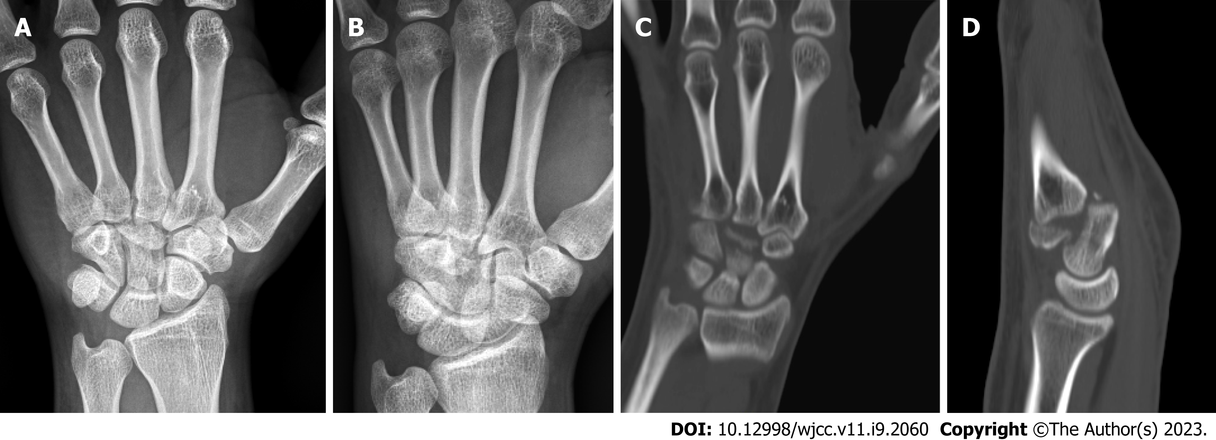
Figure 1 A fracture of the distal capitate with carpometacarpal joint dislocation is seen.
A and B: Radiography; C and D: Computed tomography of the left hand.
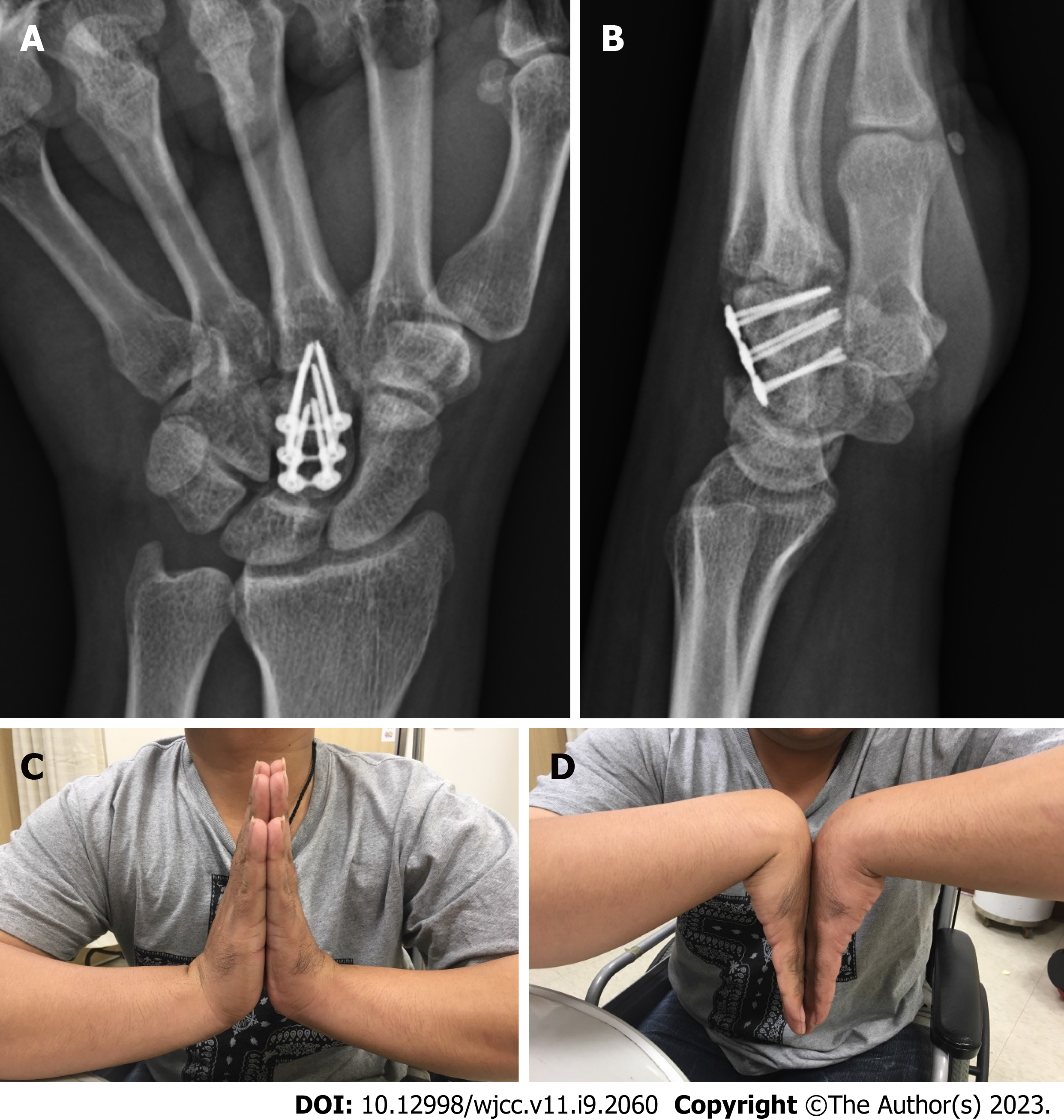
Figure 2 The flexion/extension of the left wrist is comparable to that of the dominant right wrist.
A and B: Radiography; C and D: Clinical photos of the left wrist three months postoperatively. Appropriate plate and screw placement with no fracture site collapse is observed.
Figure 3 A healed fracture with no avascular necrosis of the capitate is seen.
A and B: Radiography; C and D: Computed tomography of the left hand six years postoperatively.
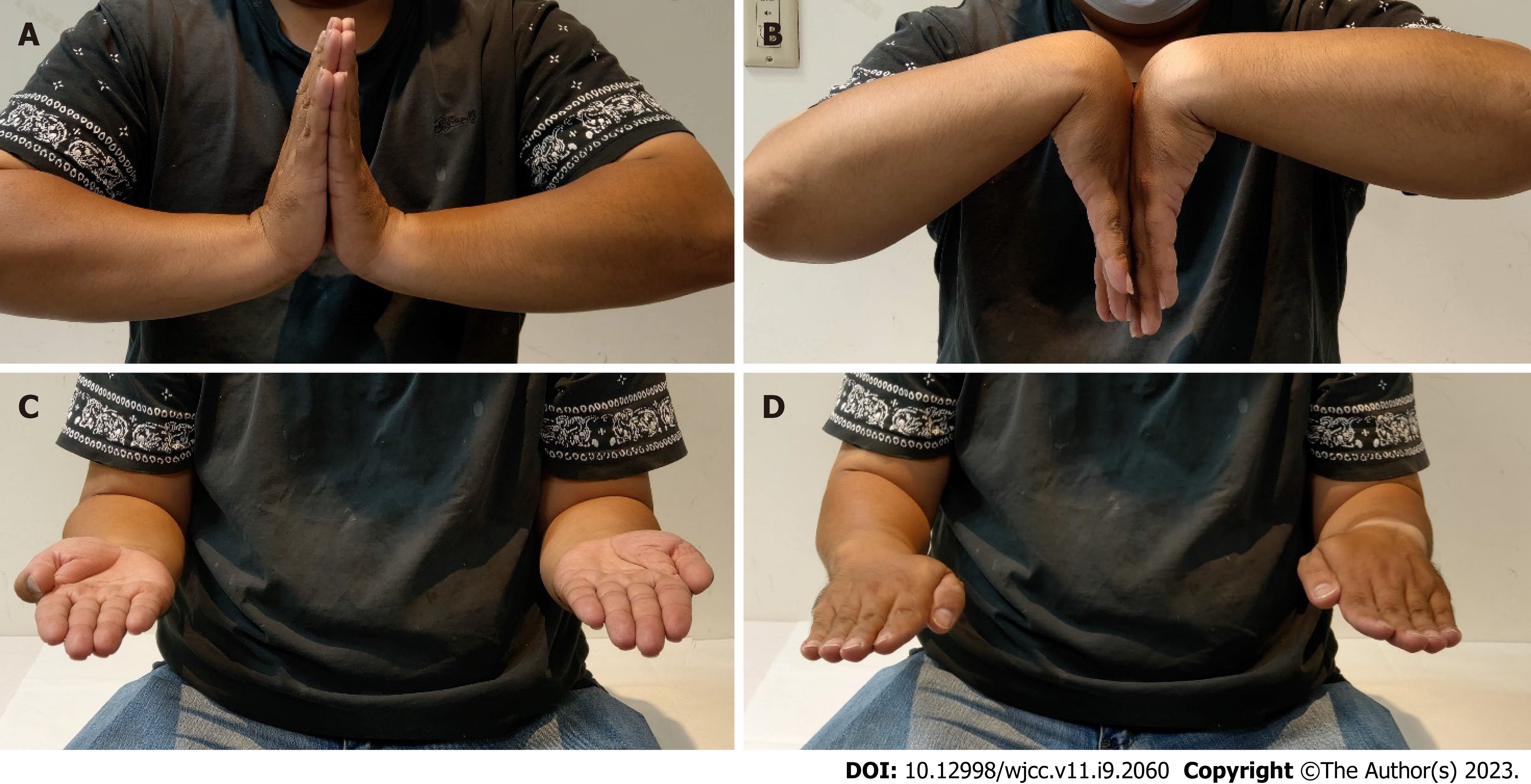
Figure 4 Clinical photos of the left wrist six years postoperatively.
A and B: The flexion/extension of left wrist; C and D: Forearm rotation are comparable to that of the dominant right wrist.
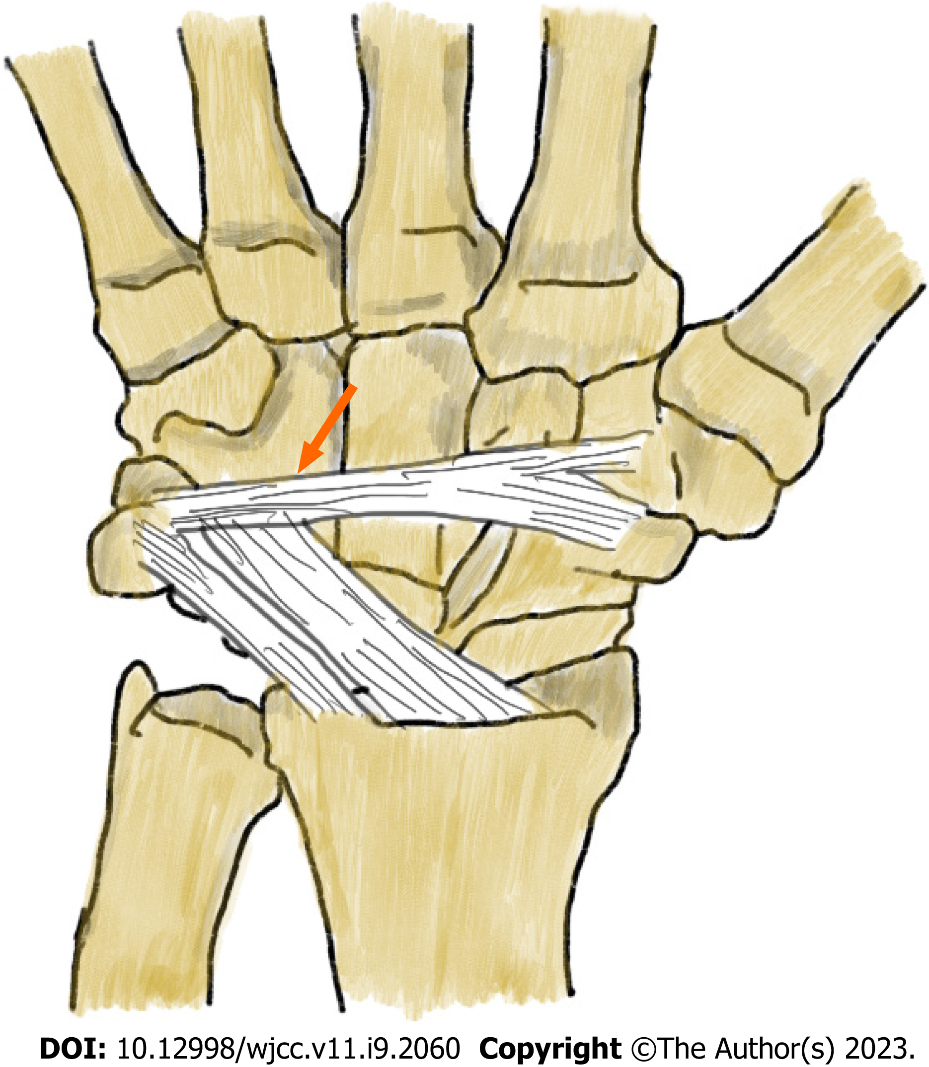
Figure 5 The proposed mechanism of injury.
The fragment is rotated by 90° because the dorsal intercarpal ligament blocks the proximal part of the fragment and due to direct axial loading on the third metacarpal base with an extremely outstretched hand.
- Citation: Lai CC, Fang HW, Chang CH, Pao JL, Chang CC, Chen YJ. Unusual capitate fracture with dorsal shearing pattern and concomitant carpometacarpal dislocation with a 6-year follow-up: A case report. World J Clin Cases 2023; 11(9): 2060-2066
- URL: https://www.wjgnet.com/2307-8960/full/v11/i9/2060.htm
- DOI: https://dx.doi.org/10.12998/wjcc.v11.i9.2060


