Copyright
©The Author(s) 2023.
World J Clin Cases. Mar 26, 2023; 11(9): 2043-2050
Published online Mar 26, 2023. doi: 10.12998/wjcc.v11.i9.2043
Published online Mar 26, 2023. doi: 10.12998/wjcc.v11.i9.2043
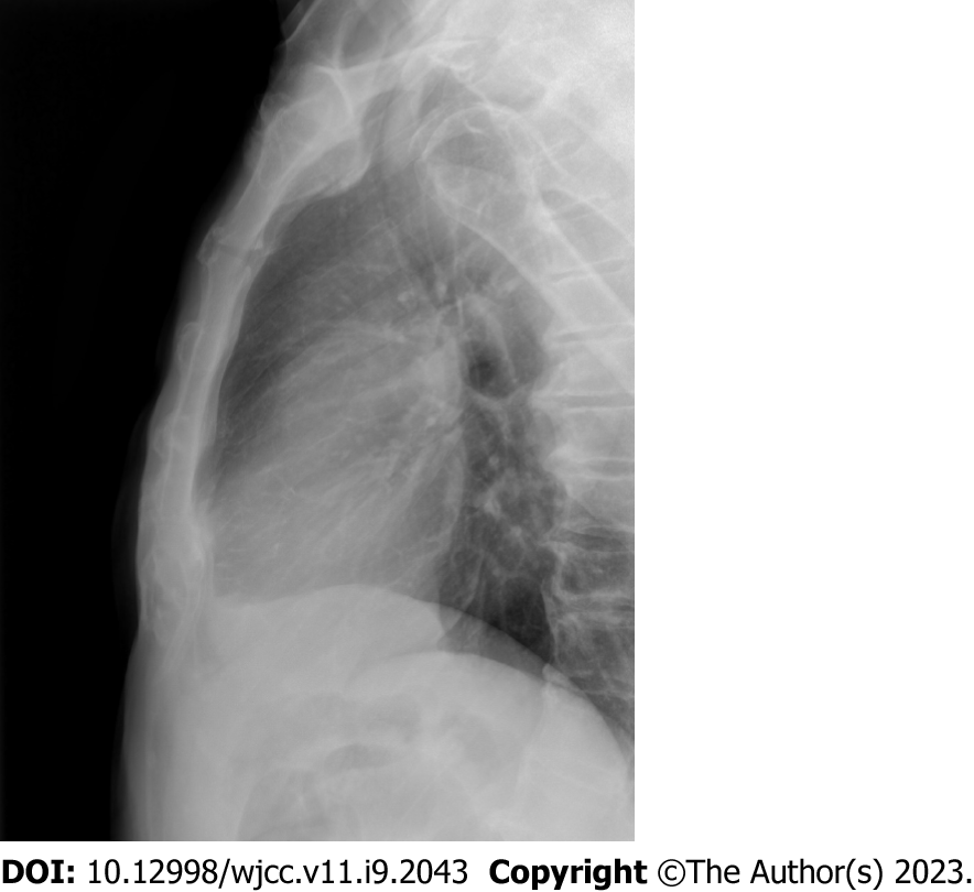
Figure 1 Lateral sternum X-ray.
The X-ray showed normal findings.
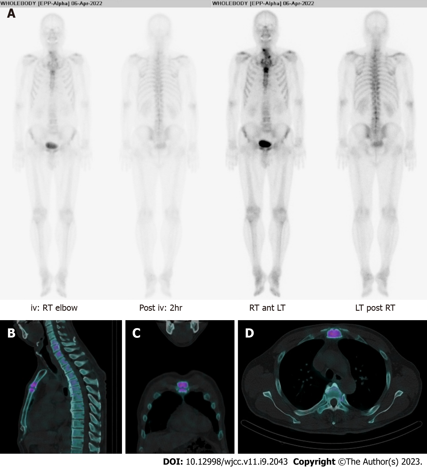
Figure 2 Bone scintigraphy and single-photon emission computed tomography-computed tomography of the manubriosternal joint.
A: Bone scintigraphy; B: Sagittal; C: Coronal; D: Axial images of single-photon emission computed tomography-computed tomography indicated arthritic change in the manubriosternal joint. iv: Intravenous; RT: Right; LT: Left.
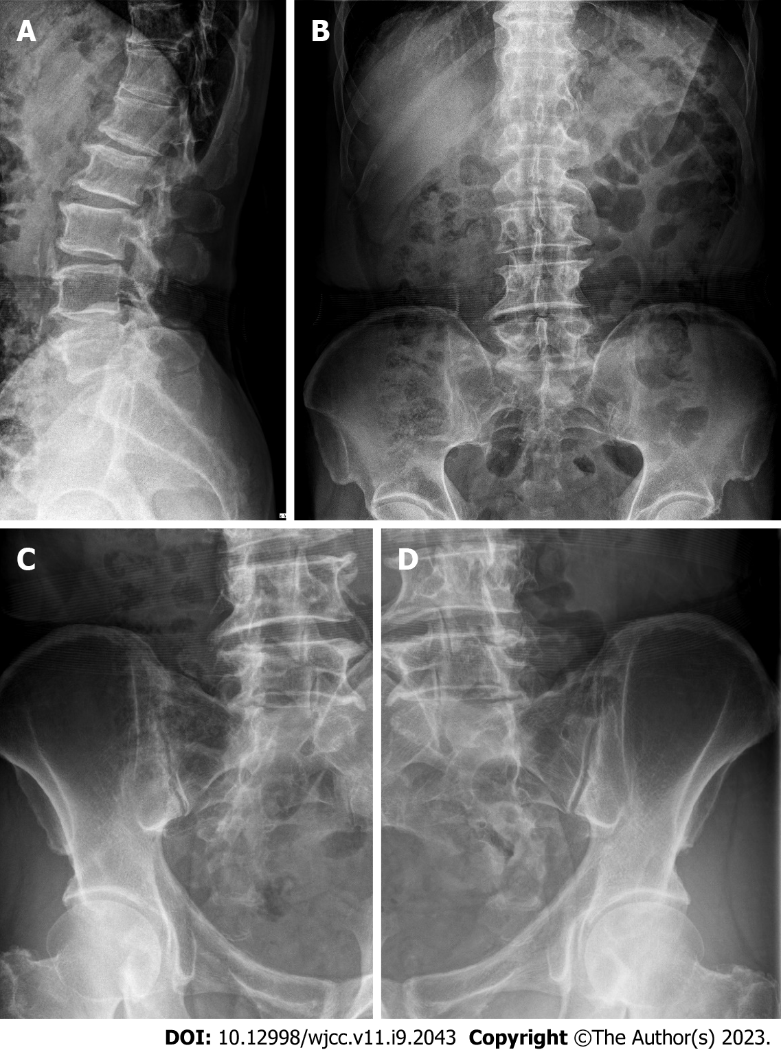
Figure 3 X-rays of the lumbar spine and sacroiliac joints.
A and B: Syndesmophytes and degenerative spondylosis were shown; A: Lateral view; B: Anterior posterior (AP) view of the lumbar spine; C and D: Bilateral sacroiliitis was found; C: AP view of the left sacroiliac joint; D: AP view of right sacroiliitis.
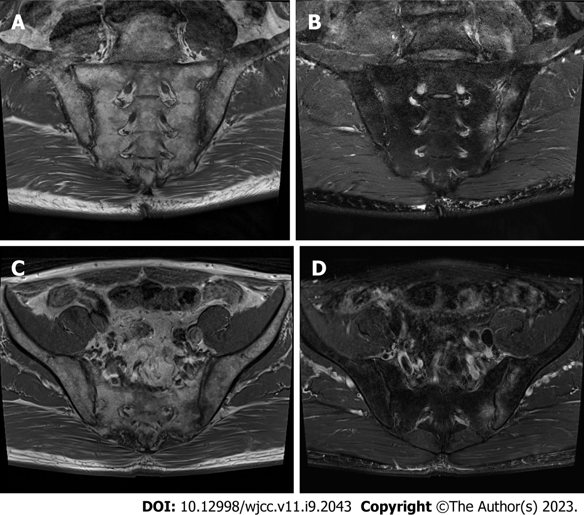
Figure 4 Magnetic resonance imaging scans of sacroiliac joints showed bilateral sacroiliitis with active inflammation at the left sacroiliac joint.
A: T1-weighted image of sacroiliac joints, coronal view; B: T2-weighted image of sacroiliac joints, coronal view; C: T1-weighted image of sacroiliac joints, axial view; D: T2-weighted image of sacroiliac joints, axial view.
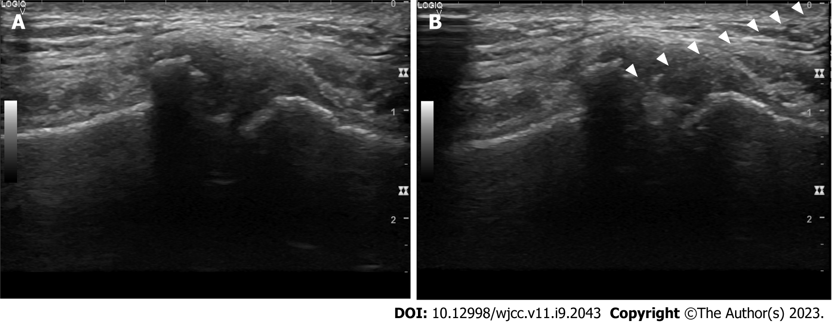
Figure 5 Ultrasound view of the manubriosternal joint.
A: Manubriosternal joint view with a 12-Hz linear probe parallel to the midsternum; B: Ultrasound-guided intra-articular corticosteroid injection into the manubriosternal joint. Arrowheads: Points at needle.
- Citation: Choi MH, Yoon IY, Kim WJ. Ultrasound-guided intra-articular corticosteroid injection in a patient with manubriosternal joint involvement of ankylosing spondylitis: A case report. World J Clin Cases 2023; 11(9): 2043-2050
- URL: https://www.wjgnet.com/2307-8960/full/v11/i9/2043.htm
- DOI: https://dx.doi.org/10.12998/wjcc.v11.i9.2043


