Copyright
©The Author(s) 2023.
World J Clin Cases. Mar 26, 2023; 11(9): 1992-2001
Published online Mar 26, 2023. doi: 10.12998/wjcc.v11.i9.1992
Published online Mar 26, 2023. doi: 10.12998/wjcc.v11.i9.1992
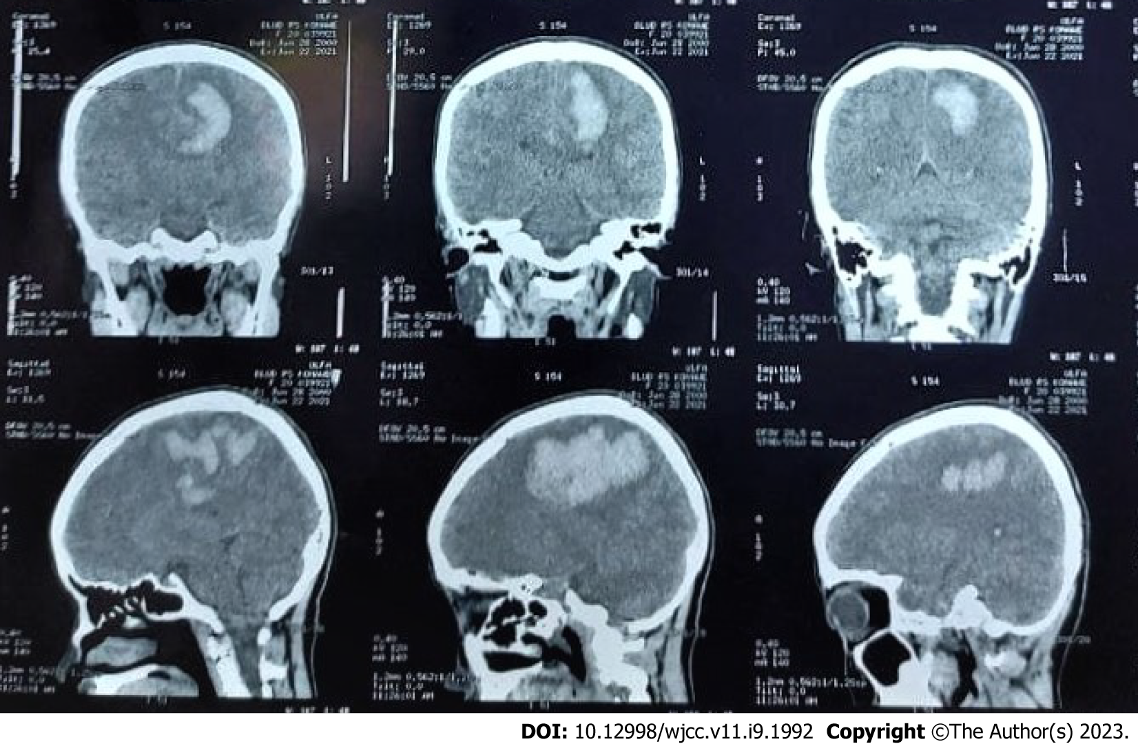
Figure 1 Head non-contrast computed tomography scan on onset (3 mo prior to presentation at our center), revealing extensive intracerebral hematoma of the left cerebral hemisphere.
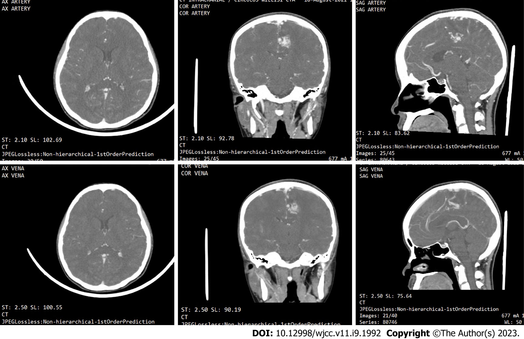
Figure 2 Subsequent head computed tomography angiography was performed 2 mo after onset, revealing the brain arteriovenous malformation, with a feeding artery from the left anterior cerebral artery, draining into the superior sagittal sinus.
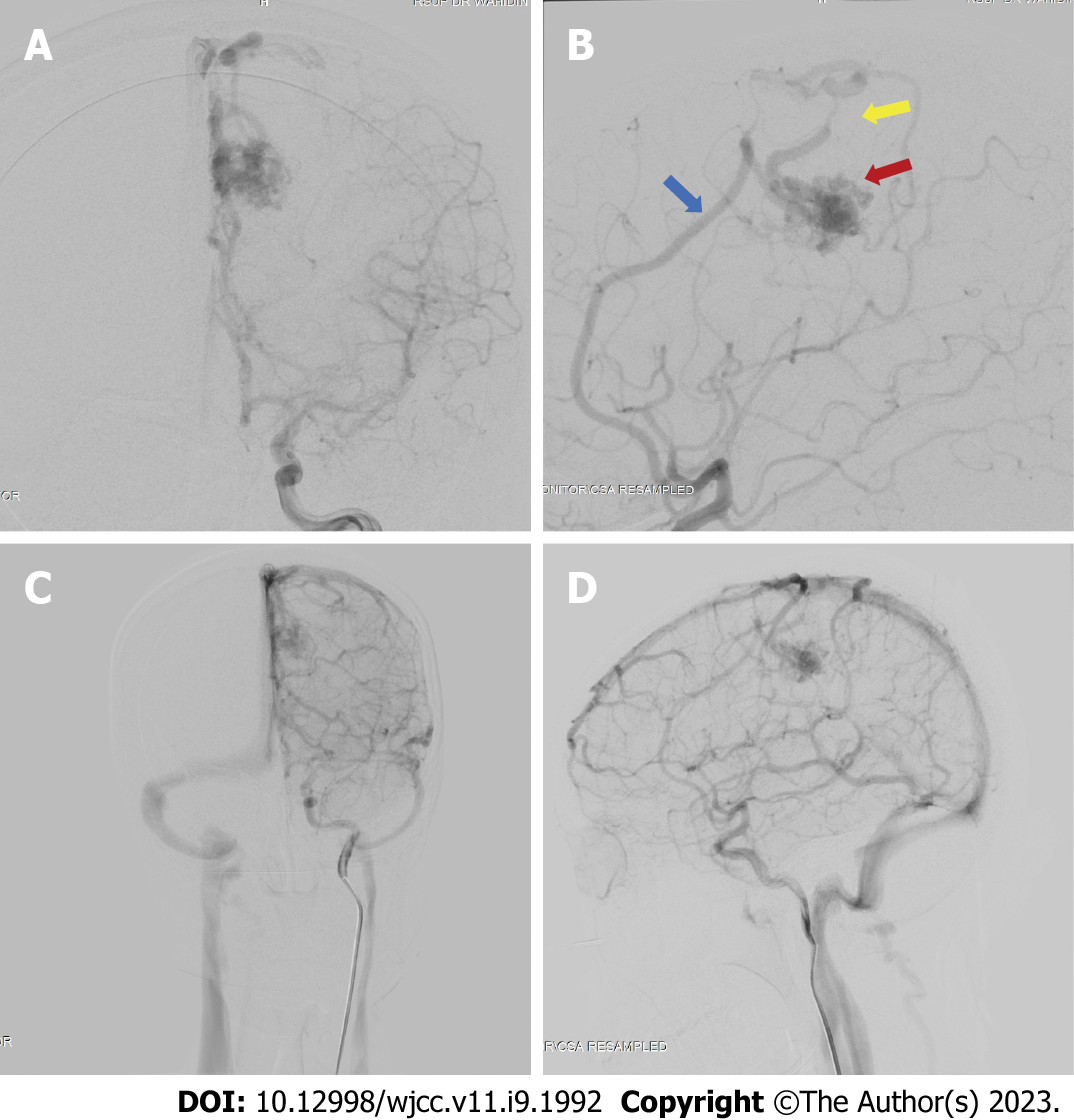
Figure 3 Digital subtraction angiography results of the patient.
The digital subtraction angiography image revealed the presence of an arteriovenous malformation (red arrow) feeding off the left pericallosal artery (blue arrow) and draining towards the cortical vein and a stenotic vein (yellow arrow). A: Late arterial phase anterior-posterior view; B: Late arterial phase, lateral view; C: Late venous phase, antero-posterior view; D: Late venous phase, lateral view.
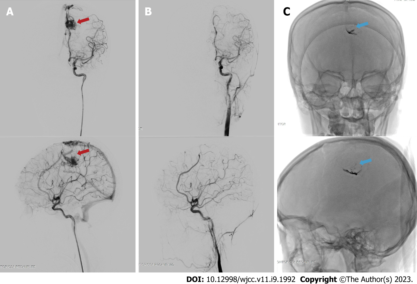
Figure 4 Digital subtraction angiography images before and after the embolization procedure on the brain arteriovenous malformation at approximately 3 mo post-ictus.
The embolization procedure was performed on the left pericallosal artery using the Onyx 18 until the nidus and draining vein can no longer be visualized. A: The nidus, before embolization (red arrow) on antero-posterior (top) and lateral (bottom) view of arterial phase; B: Complete obliteration of the nidus after embolization on the arterial phase, antero-posterior (top) and lateral (bottom) view; C: The onyx cast visible through fluoroscopy, on antero-posterior (top) and lateral (bottom) view, indicated with blue arrow.
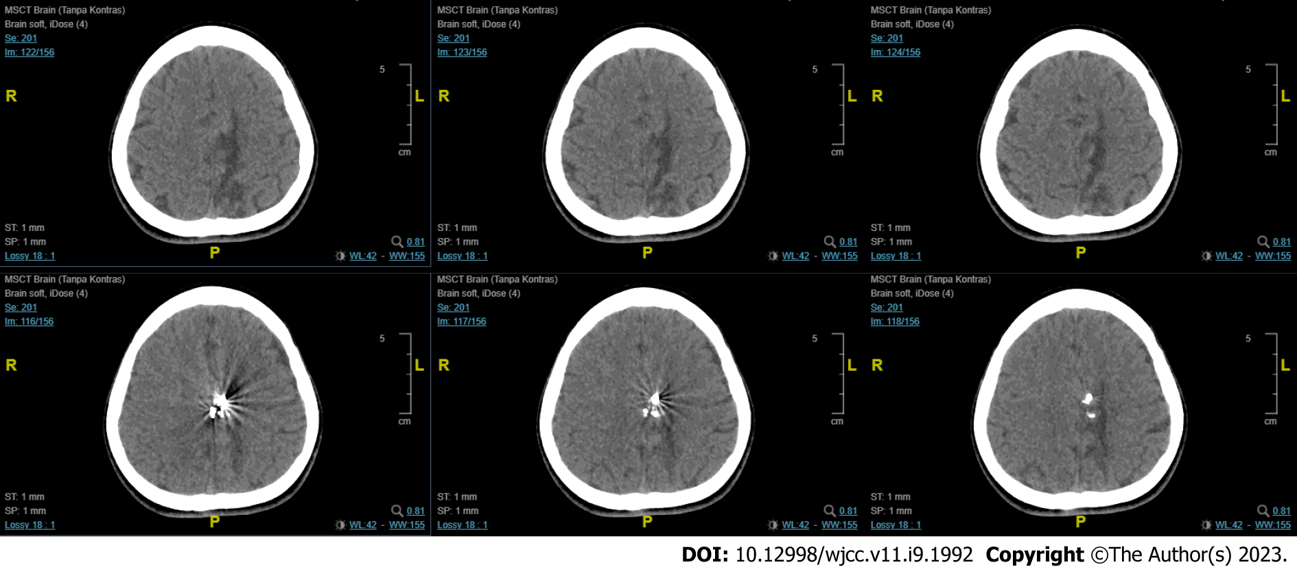
Figure 5 Follow-up computed tomography-scan revealed encephalomalacia of the parasagittal region at the left frontal and parietal lobes with mild dilatation of the left lateral ventricle.
A hyperdense blooming artefact is visualized due to the Onyx cast.
- Citation: Bintang AK, Bahar A, Akbar M, Soraya GV, Gunawan A, Hammado N, Rachman ME, Ulhaq ZS. Delayed versus immediate intervention of ruptured brain arteriovenous malformations: A case report. World J Clin Cases 2023; 11(9): 1992-2001
- URL: https://www.wjgnet.com/2307-8960/full/v11/i9/1992.htm
- DOI: https://dx.doi.org/10.12998/wjcc.v11.i9.1992


