Copyright
©The Author(s) 2023.
World J Clin Cases. Mar 16, 2023; 11(8): 1862-1868
Published online Mar 16, 2023. doi: 10.12998/wjcc.v11.i8.1862
Published online Mar 16, 2023. doi: 10.12998/wjcc.v11.i8.1862
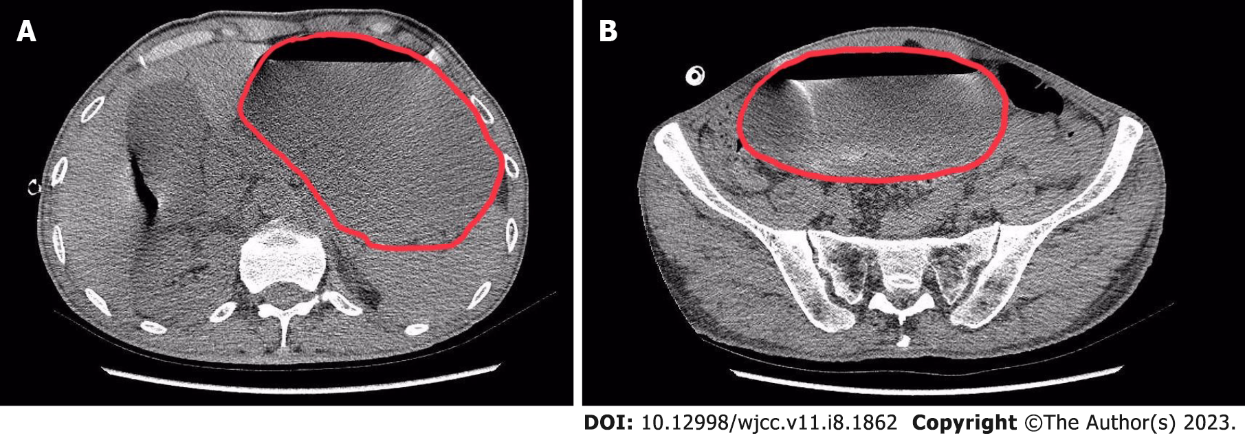
Figure 1 Computed tomography axial views of the abdominopelvic cavity 2 d after video-assisted thoracic surgery.
A: The red circle represents the scope of the stomach. In the abdominal cavity section, we can see severe distention in the stomach; B: The red circle represents the scope of the stomach. In the pelvic cavity section, we can see that the inferior edge of the stomach reached the pelvic cavity.
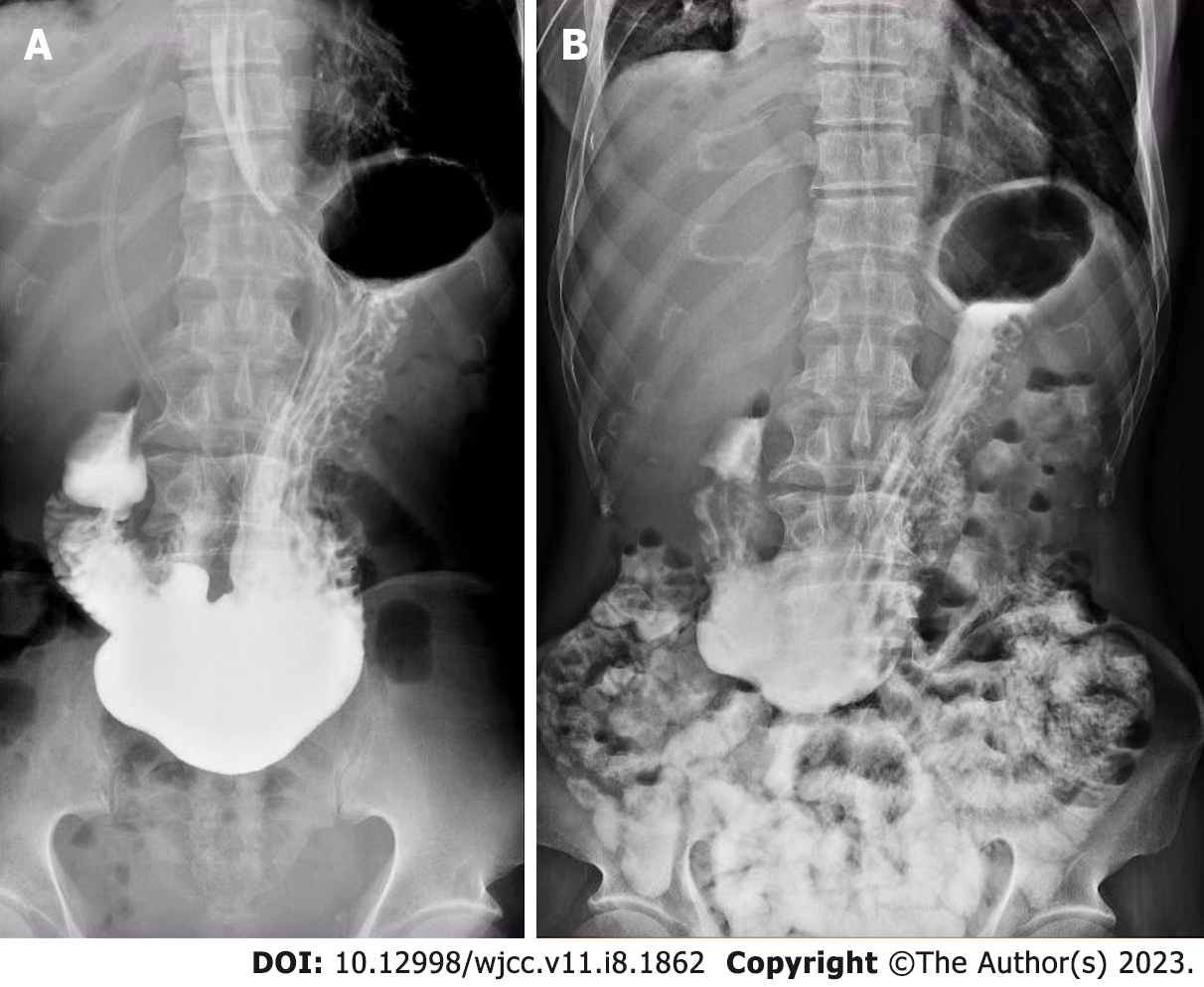
Figure 2 Oral iohexol X-ray imaging at 10 min (left) and 4 h (right) after administration of contrast agent.
A: 10 min after ingestion of iohexol, the first sign of passage to the duodenum was observed; B: Some contrast agent was retained in the stomach 4 h after administration.
- Citation: An H, Liu YC. Gastroparesis after video-assisted thoracic surgery: A case report. World J Clin Cases 2023; 11(8): 1862-1868
- URL: https://www.wjgnet.com/2307-8960/full/v11/i8/1862.htm
- DOI: https://dx.doi.org/10.12998/wjcc.v11.i8.1862


