Copyright
©The Author(s) 2023.
World J Clin Cases. Feb 26, 2023; 11(6): 1385-1392
Published online Feb 26, 2023. doi: 10.12998/wjcc.v11.i6.1385
Published online Feb 26, 2023. doi: 10.12998/wjcc.v11.i6.1385
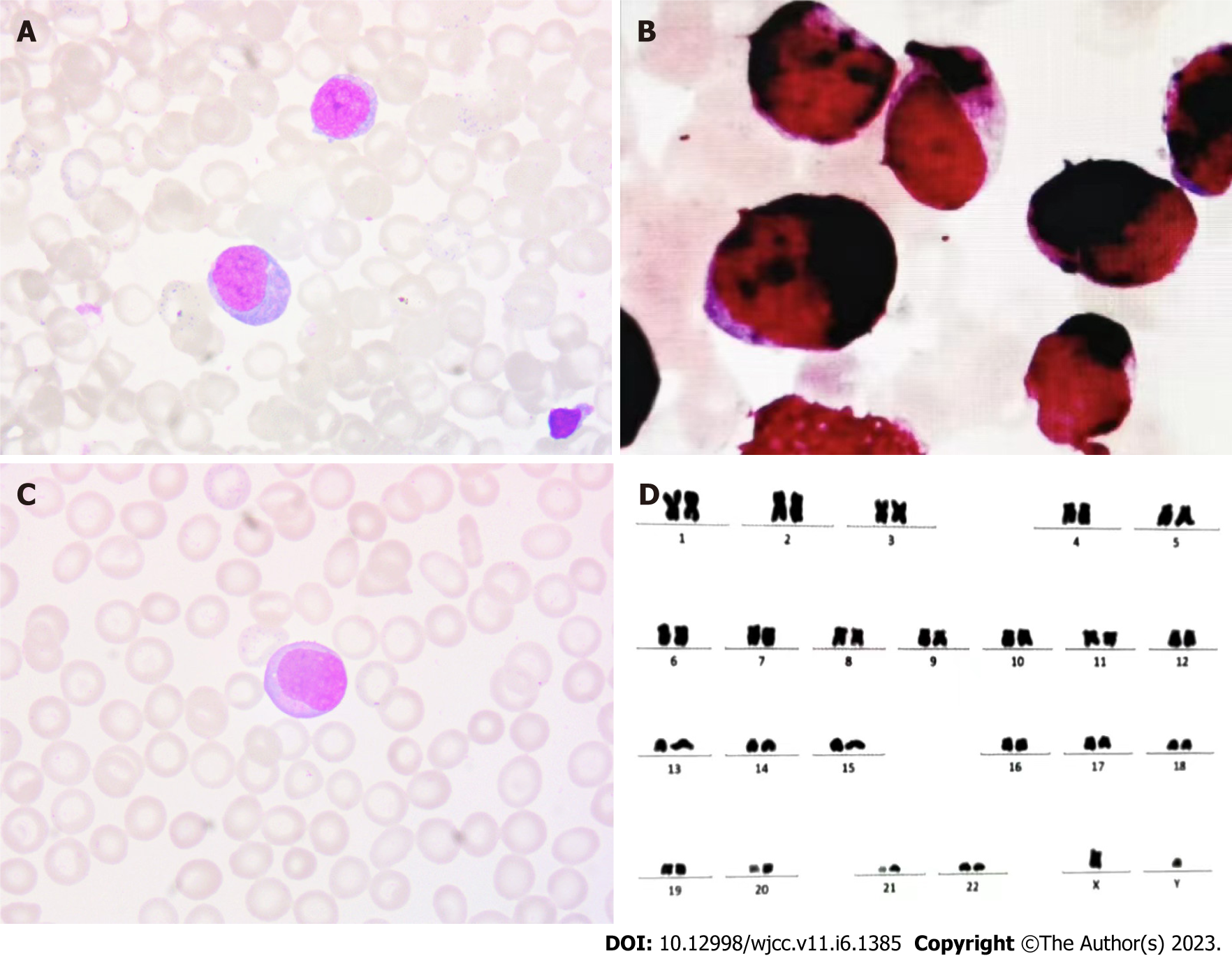
Figure 1 Chromosomal karyotyping analysis and immunohistochemical examination of the peripheral blood and bone marrow.
A: Bone marrow cell morphology revealed increased myeloblasts; B: Further immunohistochemical staining revealed an increase in myeloblasts staining strongly and weakly positively for myeloperoxidase; C: Peripheral blood imaging; D: The patient’s karyotype is 46, XY.
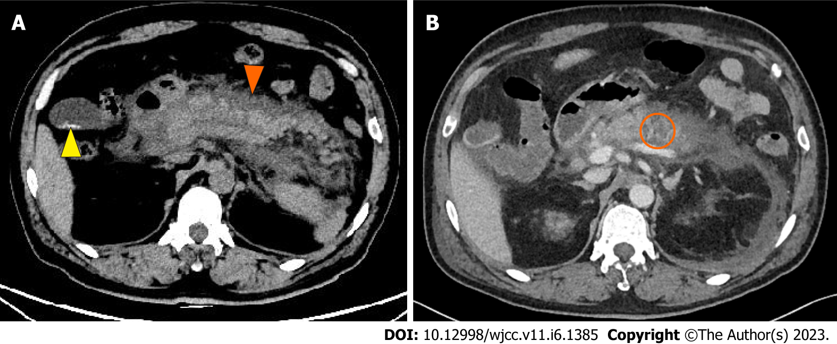
Figure 2 Imaging of infiltration of the pancreas and acute pancreatitis.
A: Abdominal computed tomography (CT) showed diffuse edema of the pancreas and peripancreatic effusion splenomegaly (orange arrow) with gallbladder stones (yellow arrow); B: Abdominal contrast-enhanced CT showed uneven density of the pancrea and no clear enhancement in the arterial phase, with hypodense lesions in the pancreas (orange circle).
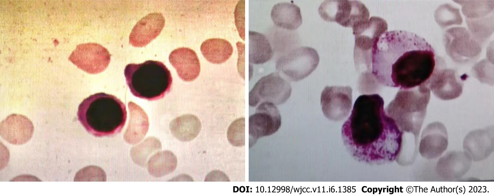
Figure 3 Bone marrow aspirate and biopsy examination.
Still active bone marrow hyperplasia and no myeloblasts indicated that hematological remission was achieved.
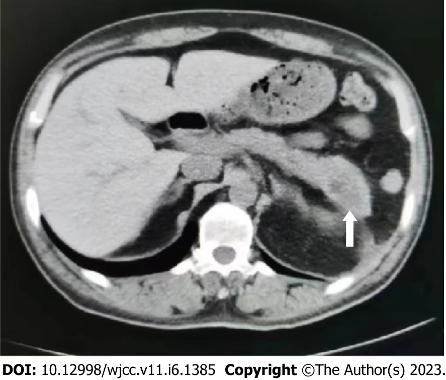
Figure 4 Imaging of the healed pancreas.
Abdominal computed tomography showed that the abnormal pancreas findings disappeared after the second course of consolidation chemotherapy, and walled-off necrosis of the pancreatic tail (white arrow).
- Citation: Yang WX, An K, Liu GF, Zhou HY, Gao JC. Acute pancreatitis as initial presentation of acute myeloid leukemia-M2 subtype: A case report. World J Clin Cases 2023; 11(6): 1385-1392
- URL: https://www.wjgnet.com/2307-8960/full/v11/i6/1385.htm
- DOI: https://dx.doi.org/10.12998/wjcc.v11.i6.1385


