Copyright
©The Author(s) 2023.
World J Clin Cases. Feb 6, 2023; 11(4): 952-961
Published online Feb 6, 2023. doi: 10.12998/wjcc.v11.i4.952
Published online Feb 6, 2023. doi: 10.12998/wjcc.v11.i4.952
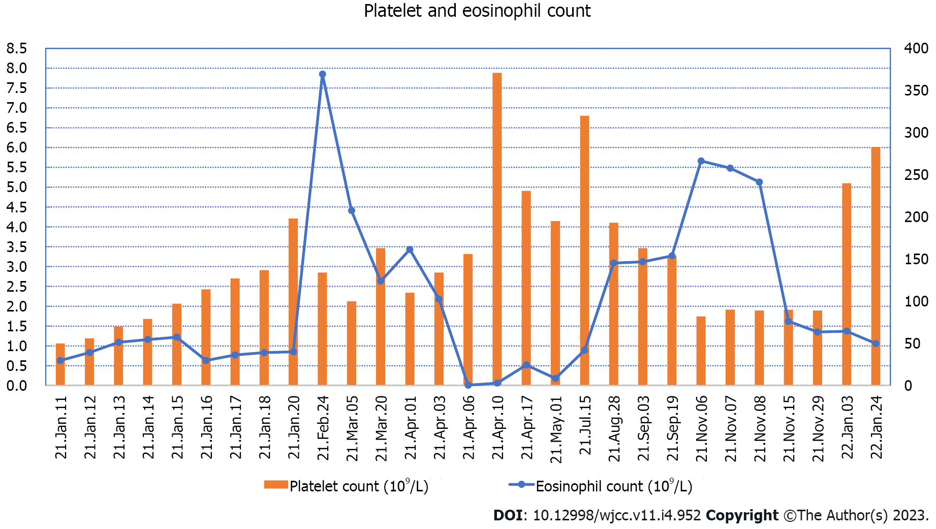
Figure 1 Changes in platelet and eosinophil count from the first hospitalization to the end of follow-up.
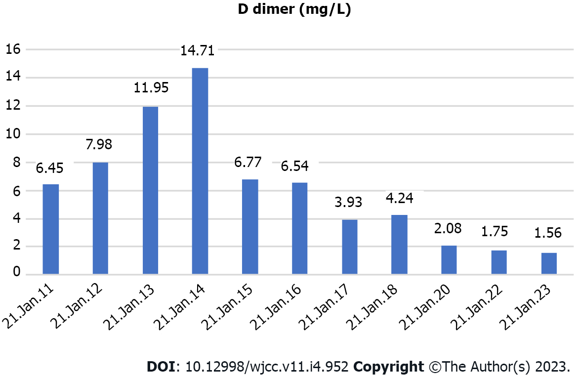
Figure 2 D dimer changes during the child's first hospitalization.
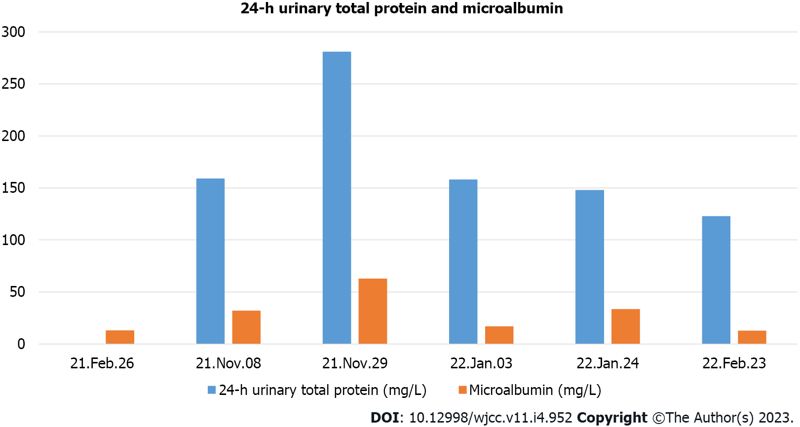
Figure 3 The 24-h urinary total protein and microalbumin levels reviewed at later stages.
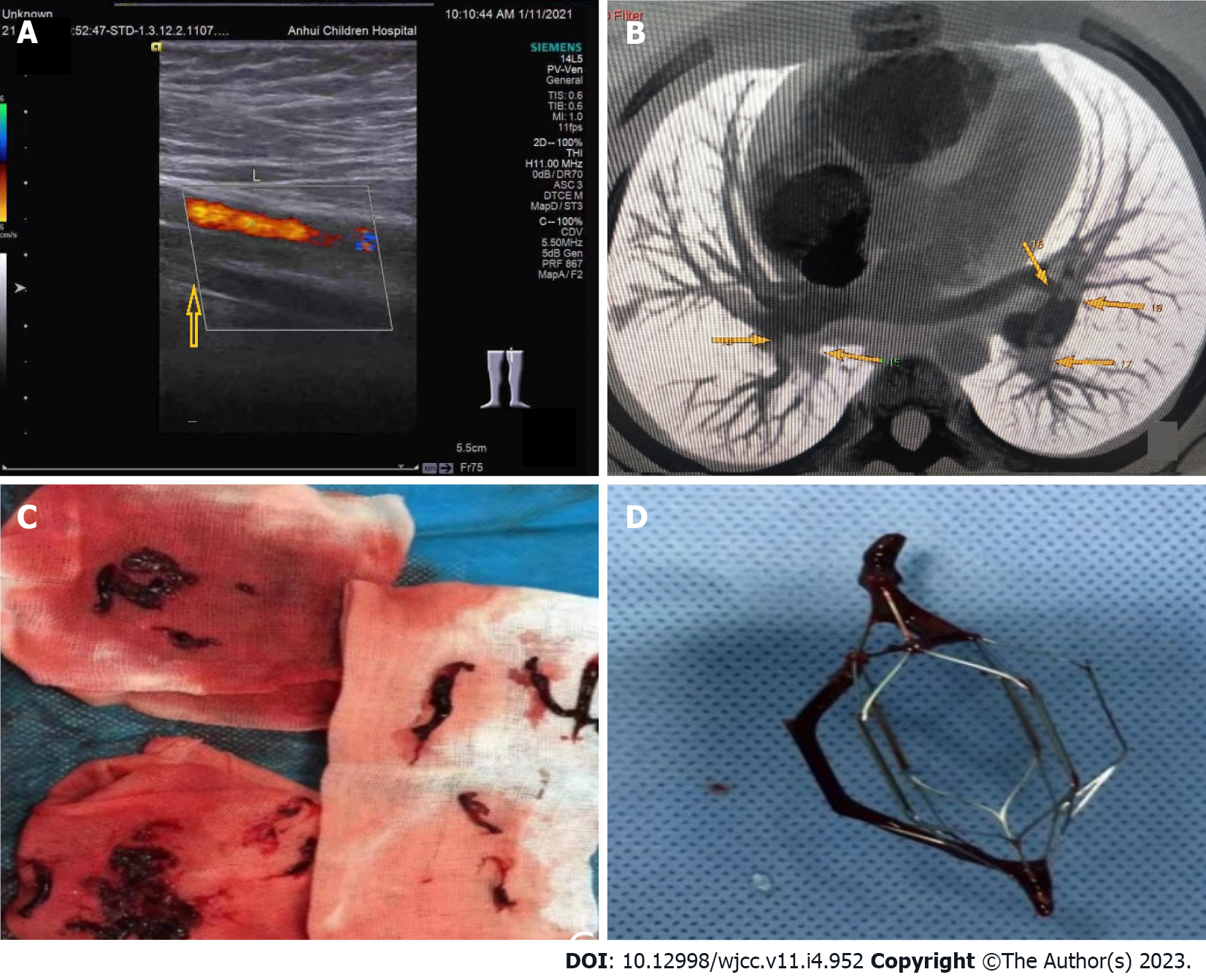
Figure 4 The 24-h urinary total protein and microalbumin levels reviewed at later stages.
A: The arrow in the figure indicates the superficial femoral vein of the left lower extremity, through which no blood flow passes. The color flow is the superficial femoral artery of the left lower extremity; B: The arrows in the figure indicate multiple filling defects in the main pulmonary arteries of both lobes and segments; C: Many long thrombi aspirated from the left leg; D: When the filter was removed, a thrombosis was attached to the filter.
- Citation: Xu YY, Huang XB, Wang YG, Zheng LY, Li M, Dai Y, Zhao S. Development of Henoch-Schoenlein purpura in a child with idiopathic hypereosinophilia syndrome with multiple thrombotic onset: A case report. World J Clin Cases 2023; 11(4): 952-961
- URL: https://www.wjgnet.com/2307-8960/full/v11/i4/952.htm
- DOI: https://dx.doi.org/10.12998/wjcc.v11.i4.952


