Copyright
©The Author(s) 2023.
World J Clin Cases. Feb 6, 2023; 11(4): 903-908
Published online Feb 6, 2023. doi: 10.12998/wjcc.v11.i4.903
Published online Feb 6, 2023. doi: 10.12998/wjcc.v11.i4.903
Figure 1 Preoperative examination and surgical planning.
A: Multiple cystic lesions with septum seen in the head and neck of the pancreas. The computer tomography value is 20 Hu. The lesions are connected to the main pancreatic duct; B: preoperative 3D reconstruction; C: plan of surgical resection with postoperative pancreatic changes are visualized in 3D reconstruction.
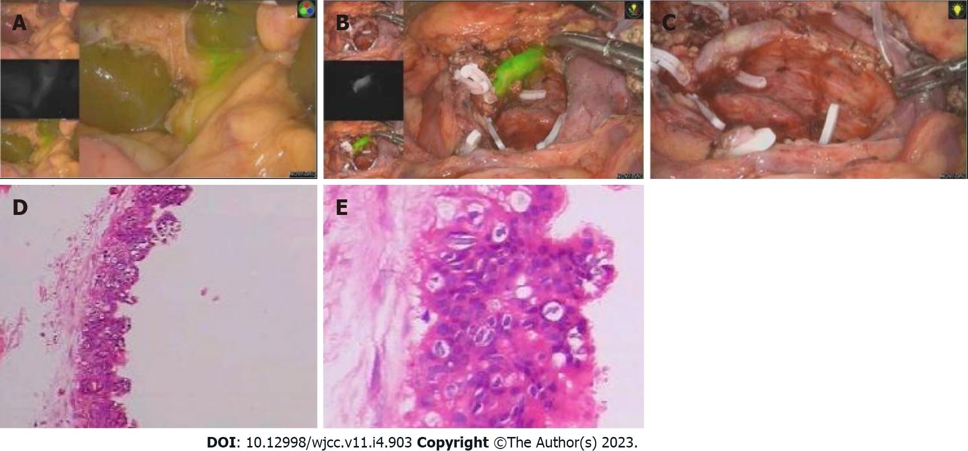
Figure 2 Intraoperative operation and postoperative pathological examination.
A: Bile duct display after 40 min of fluorescence injection; B: Surgical wound after complete resection of pancreatic head tissue under fluorescence laparoscopy; C: Surgical wound after laparoscopic complete resection of pancreatic head; D: Postoperative pathological 1; E: Postoperative pathological 2.
Figure 3 Postoperative follow-up.
A: Follow-up computer tomography (CT) after 5 mo; B: Follow-up CT after 11 mo; C: Follow-up CT after 17 mo.
- Citation: Li XL, Gong LS. Preoperative 3D reconstruction and fluorescent indocyanine green for laparoscopic duodenum preserving pancreatic head resection: A case report. World J Clin Cases 2023; 11(4): 903-908
- URL: https://www.wjgnet.com/2307-8960/full/v11/i4/903.htm
- DOI: https://dx.doi.org/10.12998/wjcc.v11.i4.903


