Copyright
©The Author(s) 2023.
World J Clin Cases. Dec 26, 2023; 11(36): 8581-8588
Published online Dec 26, 2023. doi: 10.12998/wjcc.v11.i36.8581
Published online Dec 26, 2023. doi: 10.12998/wjcc.v11.i36.8581
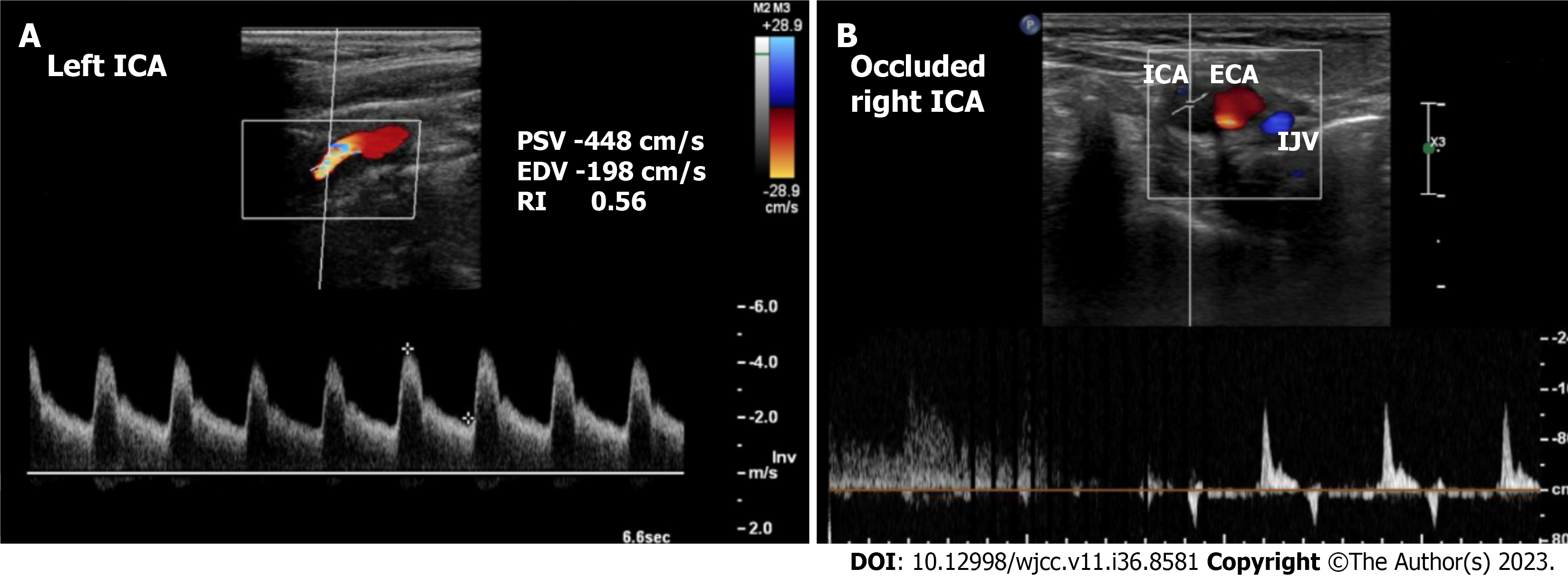
Figure 1 Preoperative carotid ultrasound duplex in case 1.
A: Left internal carotid artery stenosis of > 90%; B: Totally occluded right interval carotid artery. ECA: External carotid artery; EDV: End-diastolic velocity; IJV: Internal jugular vein; PSV: Peak systolic velocity; RI: Resistance index; ICA: Internal carotid artery.
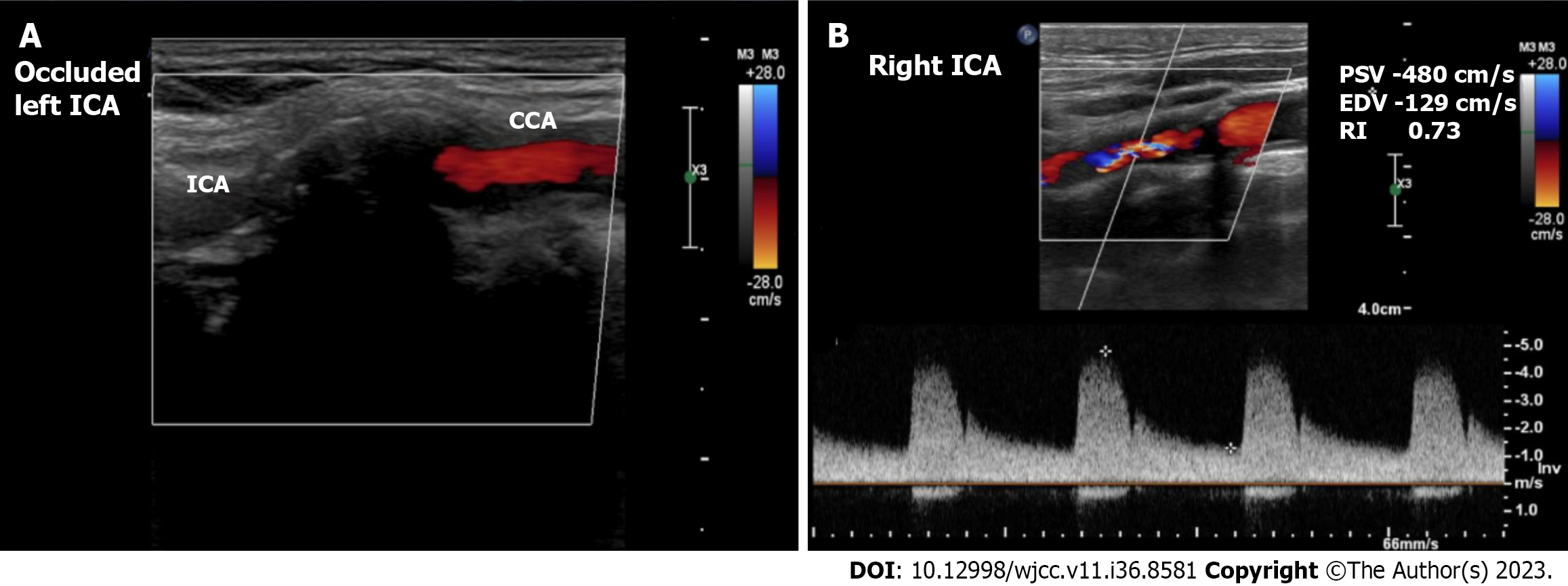
Figure 2 Preoperative carotid ultrasound duplex in case 2.
A: Totally occluded left internal carotid artery; B: Right interval carotid artery stenosis of > 90%. CCA: Common carotid artery; EDV: End-diastolic velocity; PSV: Peak systolic velocity; RI: Resistance index; ICA: Internal carotid artery.
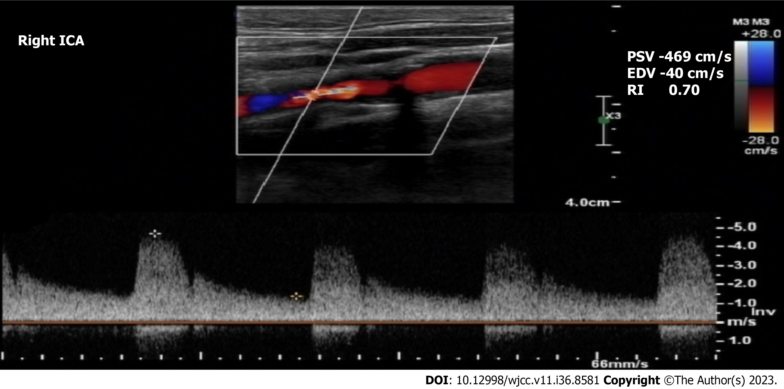
Figure 3 Preoperative carotid ultrasound duplex showed right internal carotid artery stenosis of > 90% in case 3.
EDV: End-diastolic velocity; PSV: Peak systolic velocity; RI: Resistance index; ICA: Internal carotid artery.
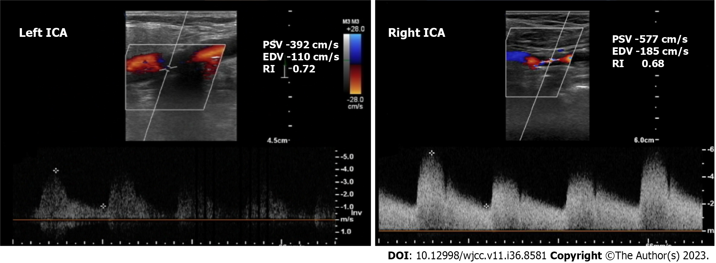
Figure 4 Preoperative carotid ultrasound duplex in case 4.
Bilateral internal carotid artery stenosis of > 90%. EDV: End-diastolic velocity; PSV: Peak systolic velocity; RI: Resistance index; ICA: Internal carotid artery.
- Citation: AlGhamdi FK, Altoijry A, AlQahtani A, Aldossary MY, AlSheikh SO, Iqbal K, Alayadhi WA. Synchronous carotid endarterectomy and coronary artery bypass graft: Four case reports. World J Clin Cases 2023; 11(36): 8581-8588
- URL: https://www.wjgnet.com/2307-8960/full/v11/i36/8581.htm
- DOI: https://dx.doi.org/10.12998/wjcc.v11.i36.8581


