Copyright
©The Author(s) 2023.
World J Clin Cases. Dec 26, 2023; 11(36): 8568-8573
Published online Dec 26, 2023. doi: 10.12998/wjcc.v11.i36.8568
Published online Dec 26, 2023. doi: 10.12998/wjcc.v11.i36.8568
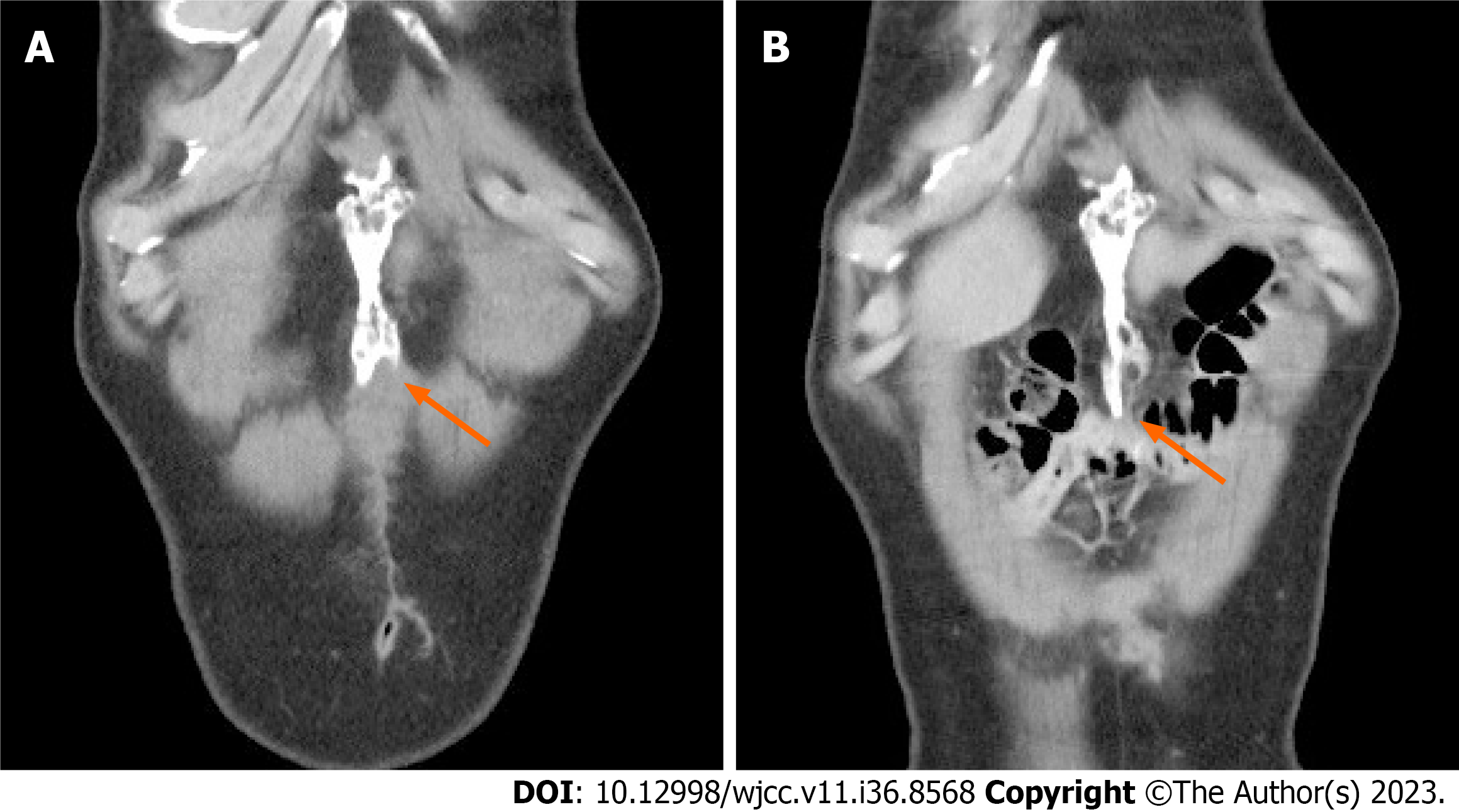
Figure 1 Computerized tomography images showing heterotopic ossification (arrow).
A: A 10 cm-sized ossification was located along the laparotomy scar six months after surgery; B: A slight increase in size was observed one year after surgery.
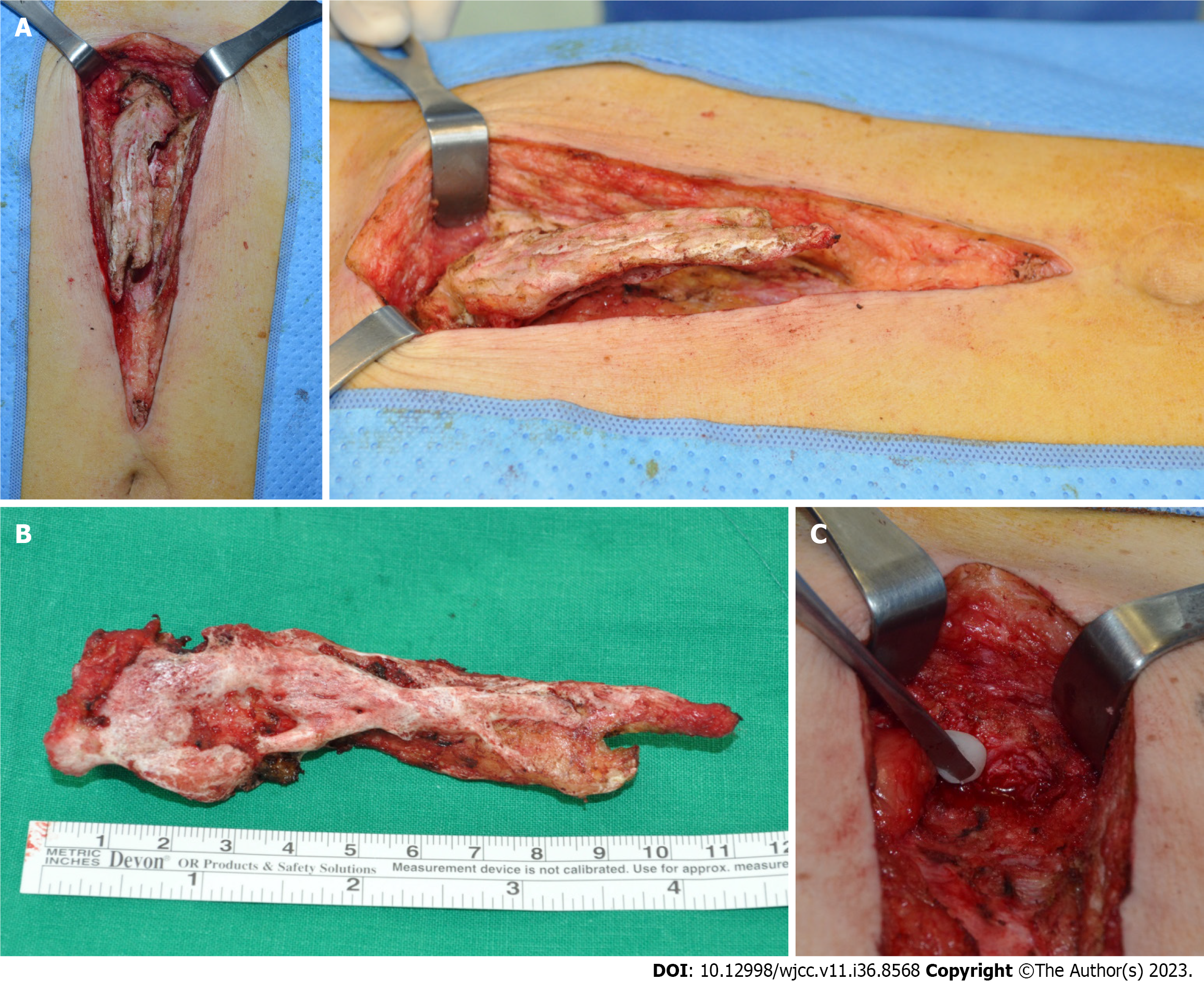
Figure 2 Excision of heterotopic ossification.
A: The structure originated from the xiphoid process, extending inferiorly along the midline of the abdomen; B: The 11 cm-sized ossification was excised completely; C: Bone wax was applied to the excisional margin.
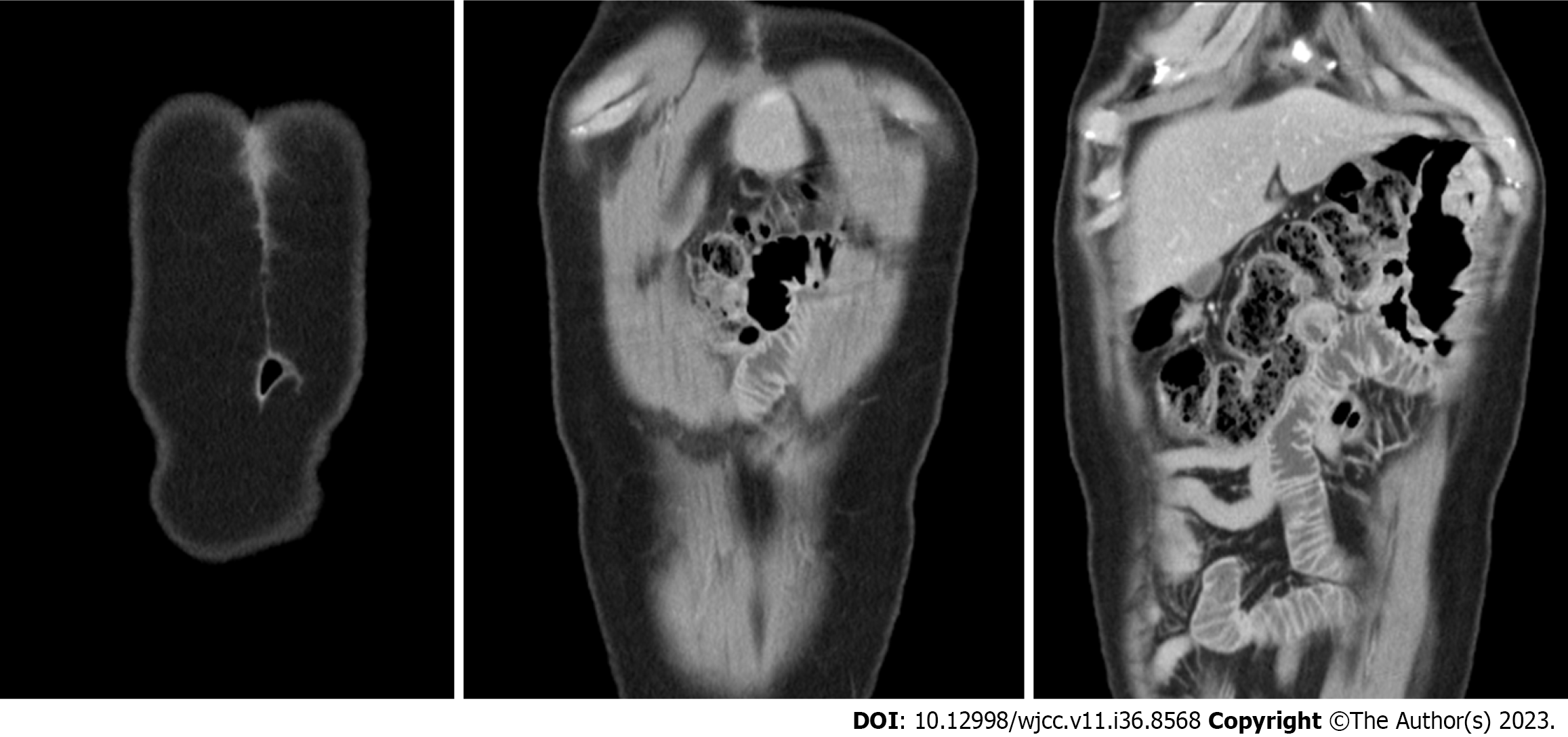
Figure 3 An abdominal computerized tomographic scan, taken one year after excision, showing no sign of recurred heterotopic ossification.
- Citation: Lee SS. Post-laparotomy heterotopic ossification of the xiphoid process: A case report. World J Clin Cases 2023; 11(36): 8568-8573
- URL: https://www.wjgnet.com/2307-8960/full/v11/i36/8568.htm
- DOI: https://dx.doi.org/10.12998/wjcc.v11.i36.8568


