Copyright
©The Author(s) 2023.
World J Clin Cases. Dec 26, 2023; 11(36): 8563-8567
Published online Dec 26, 2023. doi: 10.12998/wjcc.v11.i36.8563
Published online Dec 26, 2023. doi: 10.12998/wjcc.v11.i36.8563
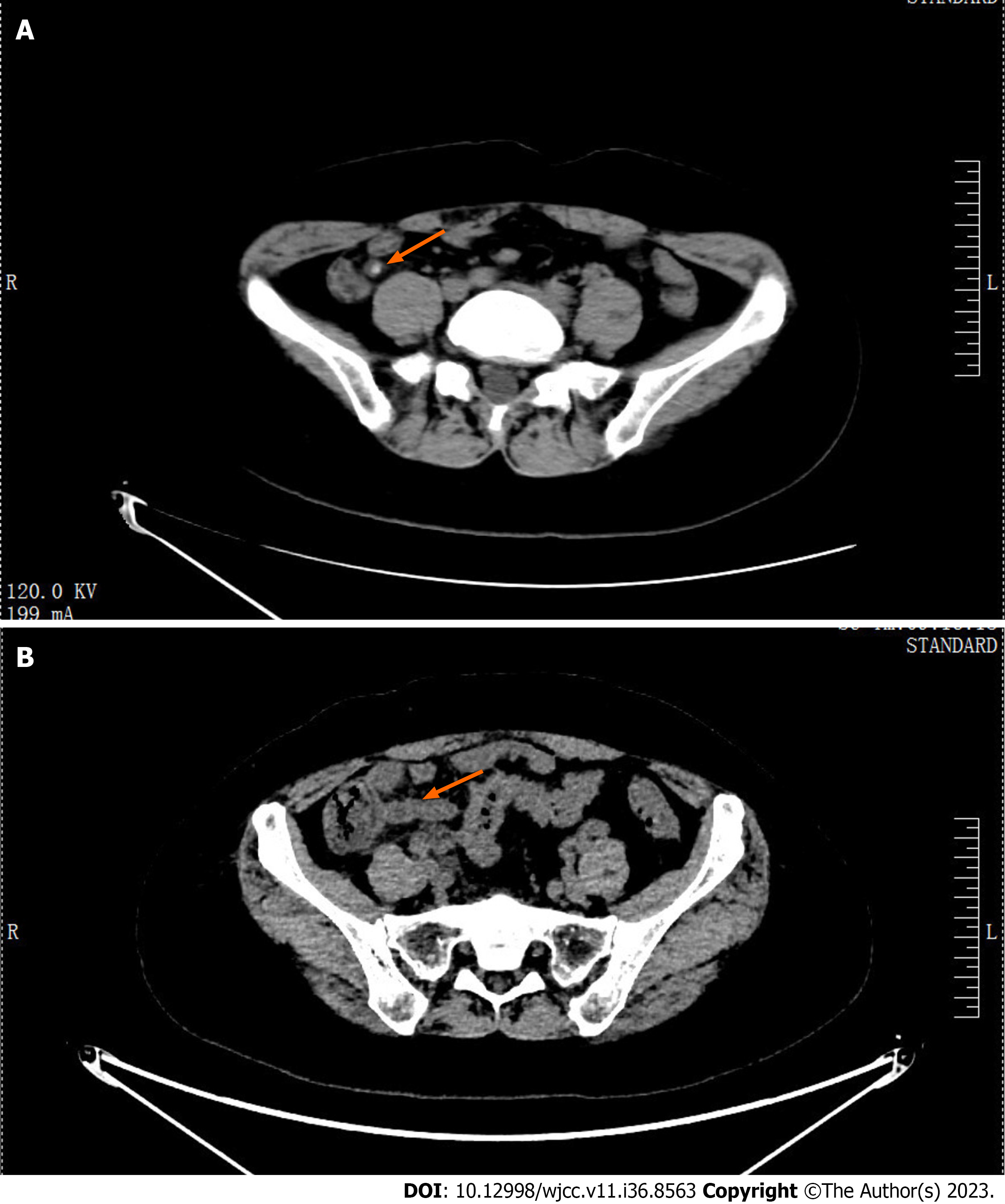
Figure 1 Computed tomography scan of the lower abdomen and pelvis.
A: Computed tomography (CT) revealed a dilated and thickened appendix with fecoliths (solid arrow: Appendix with fecoliths); B: After 3 d of treatment, the pelvic CT revealed that the appendicolith had disappeared (solid arrow: Dilated appendix without fecolith).
- Citation: Song XL, Ma JY, Zhang ZG. Colonoscopy-induced acute appendicitis: A case report. World J Clin Cases 2023; 11(36): 8563-8567
- URL: https://www.wjgnet.com/2307-8960/full/v11/i36/8563.htm
- DOI: https://dx.doi.org/10.12998/wjcc.v11.i36.8563


