Copyright
©The Author(s) 2023.
World J Clin Cases. Dec 26, 2023; 11(36): 8557-8562
Published online Dec 26, 2023. doi: 10.12998/wjcc.v11.i36.8557
Published online Dec 26, 2023. doi: 10.12998/wjcc.v11.i36.8557
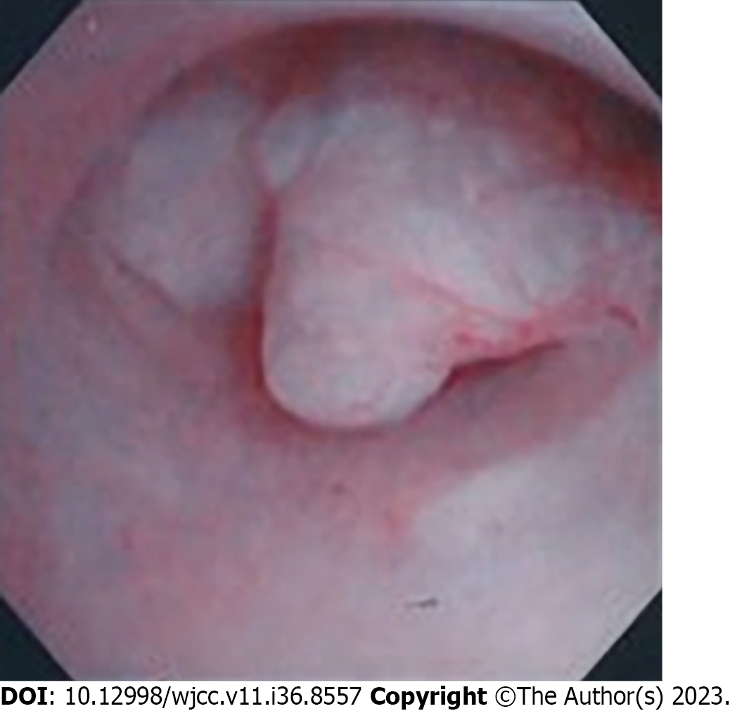
Figure 1 Hysteroscopic findings.
A pale-red, elevated lesion is observed in the lower portion of the uterine body.
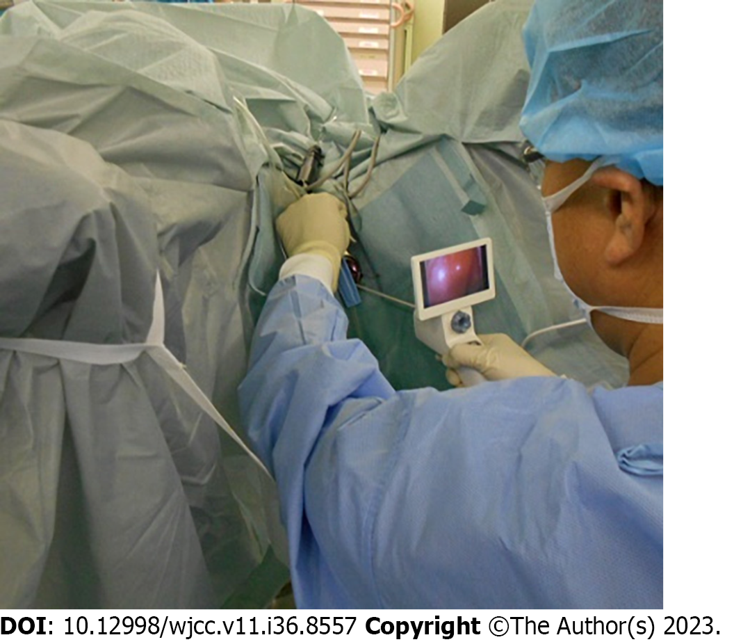
Figure 2 Surgical procedure.
The uterine lumen is observed using a LiNA OperaScopeTM device.
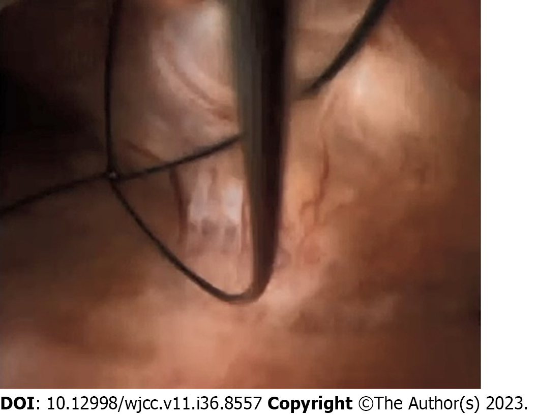
Figure 3 Performance of endometrial polypectomy.
Endometrial polyps are removed using basket forceps while under LiNA OperaScopeTM endoscopy.
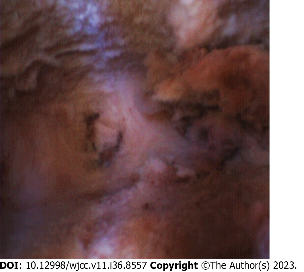
Figure 4 Post-microwave endometrial ablation hysteroscopic findings.
The absence of uncauterized areas of the endometrium and necrotic tissue following endometrial cauterization is confirmed.
- Citation: Kakinuma K, Kakinuma T, Ueyama K, Shinohara T, Okamoto R, Yanagida K, Takeshima N, Ohwada M. LiNA OperaScopeTM for microwave endometrial ablation for endometrial polyps with heavy menstrual bleeding: A case report. World J Clin Cases 2023; 11(36): 8557-8562
- URL: https://www.wjgnet.com/2307-8960/full/v11/i36/8557.htm
- DOI: https://dx.doi.org/10.12998/wjcc.v11.i36.8557


