Copyright
©The Author(s) 2023.
World J Clin Cases. Dec 26, 2023; 11(36): 8519-8526
Published online Dec 26, 2023. doi: 10.12998/wjcc.v11.i36.8519
Published online Dec 26, 2023. doi: 10.12998/wjcc.v11.i36.8519
Figure 1 Abdominopelvic computed tomography.
Coronal contrast-enhanced abdominal computed tomography reveals a contracted gallbladder (white arrowhead) in close contact with the second portion of the duodenum (orange arrowhead), with a compromised fat plane between these two structures (white dotted line). A soft-tissue density (white arrow) connects the contracted gallbladder and transverse colon (asterisk).
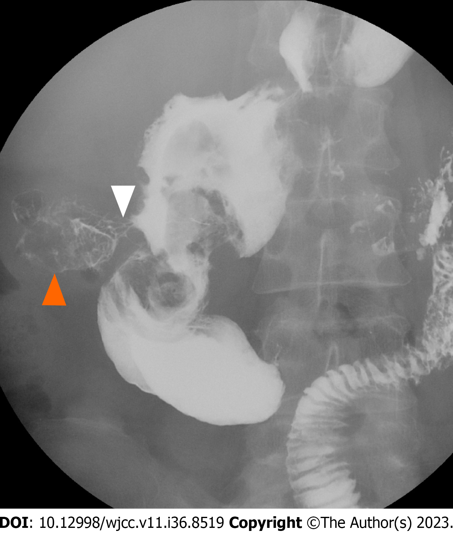
Figure 2 Barium upper gastrointestinal series.
The examination reveals a contrast-filling sac-like structure in the right lower quadrant of the abdomen (orange arrowhead) connected to the second portion of the duodenum (white arrowhead).
- Citation: Wang CY, Chiu SH, Chang WC, Ho MH, Chang PY. Cholecystoenteric fistula in a patient with advanced gallbladder cancer: A case report and review of literature. World J Clin Cases 2023; 11(36): 8519-8526
- URL: https://www.wjgnet.com/2307-8960/full/v11/i36/8519.htm
- DOI: https://dx.doi.org/10.12998/wjcc.v11.i36.8519


