Copyright
©The Author(s) 2023.
World J Clin Cases. Dec 26, 2023; 11(36): 8512-8518
Published online Dec 26, 2023. doi: 10.12998/wjcc.v11.i36.8512
Published online Dec 26, 2023. doi: 10.12998/wjcc.v11.i36.8512
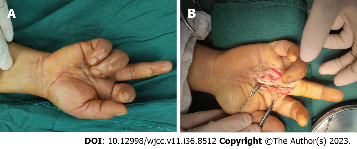
Figure 1 Findings during the first surgery.
A: The extended middle finger and flexed ring finger before exploration; B: Intraoperative view of the rupture of both flexor digitorum superficialis and flexor digitorum profundus tendon at the metacarpophalangeal joint of the middle finger.
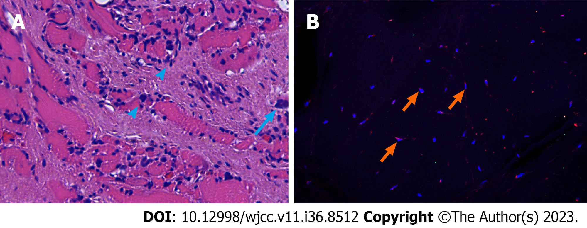
Figure 2 Results of hematoxylin and eosin staining and immunofluorescence staining of the flexor digitorum profundus muscle.
A: Interstitial diffuse infiltration of scattered lymphocytes (arrowheads) and multinucleated giant cells (arrow) in the muscle tissue; B: The cells were stained for nuclei (DAPI, blue) and CD68 (red). This highlights the giant cells that are positively immunostained for CD68 (arrows).
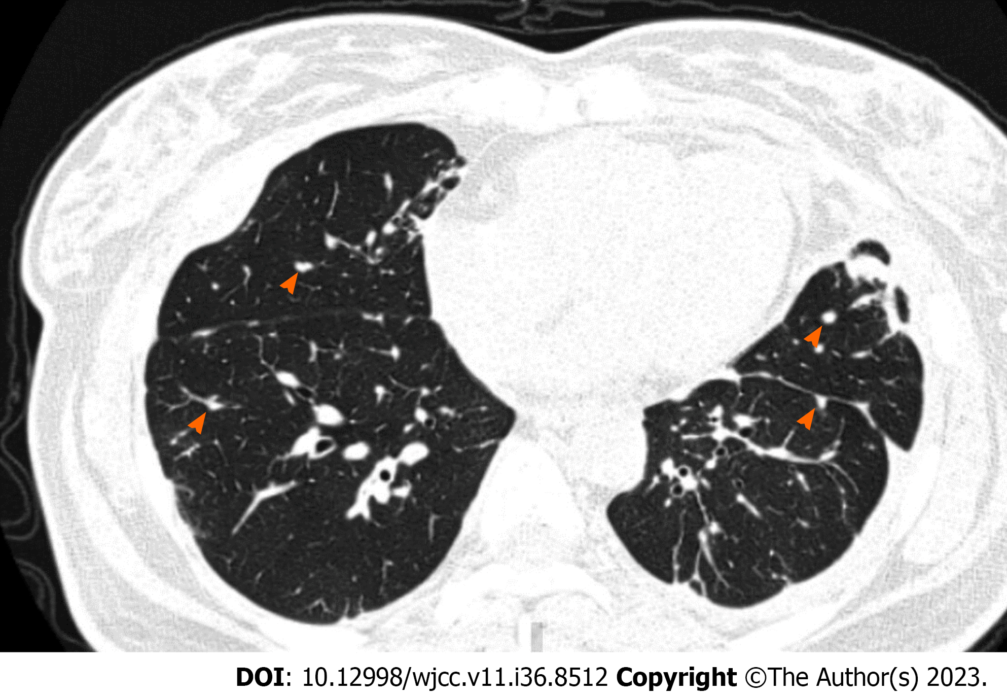
Figure 3
High-resolution computed tomographic section showing widespread nodules (arrowheads) in both lungs.
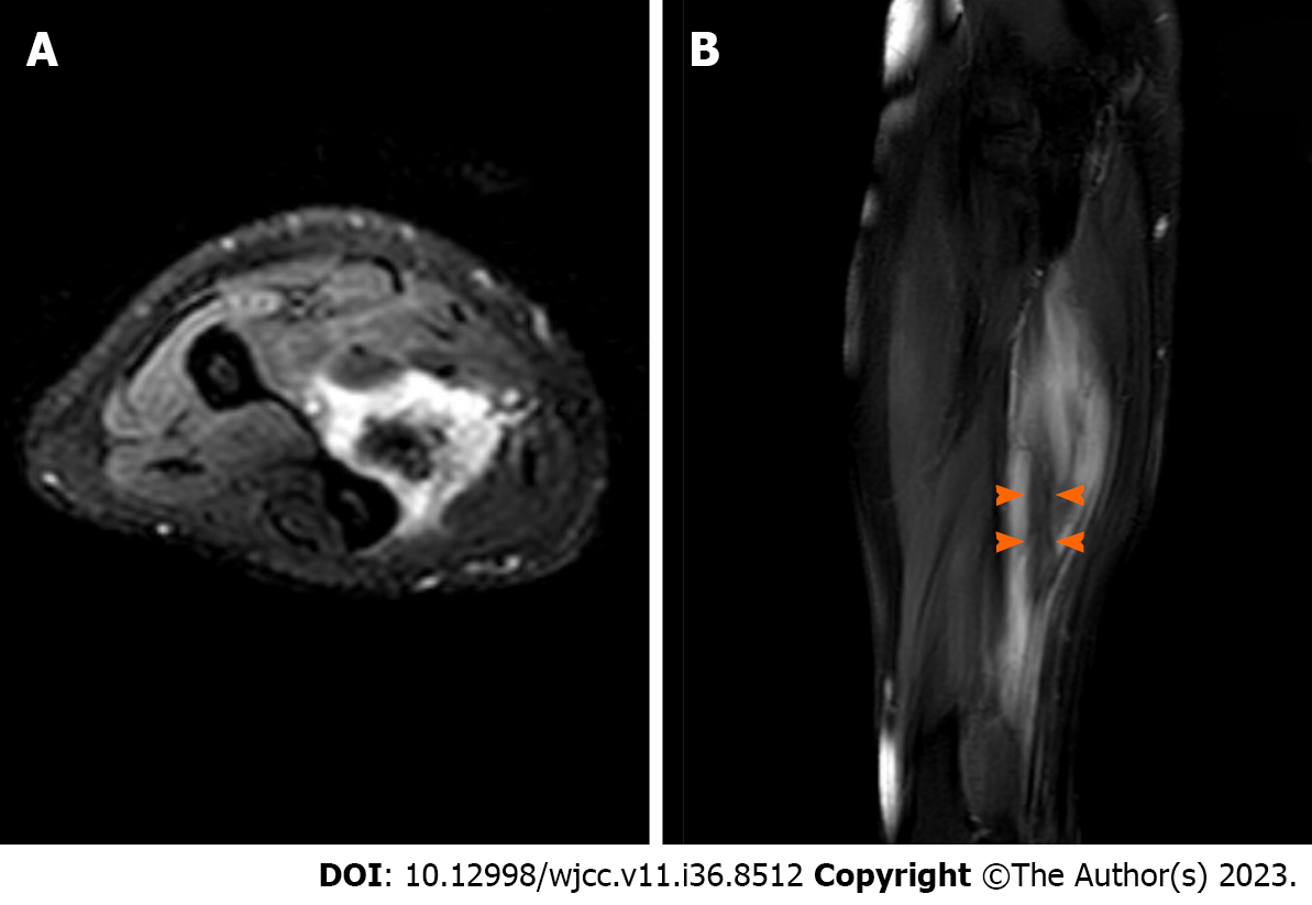
Figure 4 Magnetic resonance imaging revealing a lesion located at the flexor digitorum profundus muscle.
A: T2-weighted fat saturated axial image of the forearm shows a nodule of increased signal intensity, while the center structure shows decreased signal intensity; B: T2-weighted fat saturated sagittal image of the forearm shows an inner stripe of decreased signal intensity (arrowheads) and outer stripes of increased signal intensity.
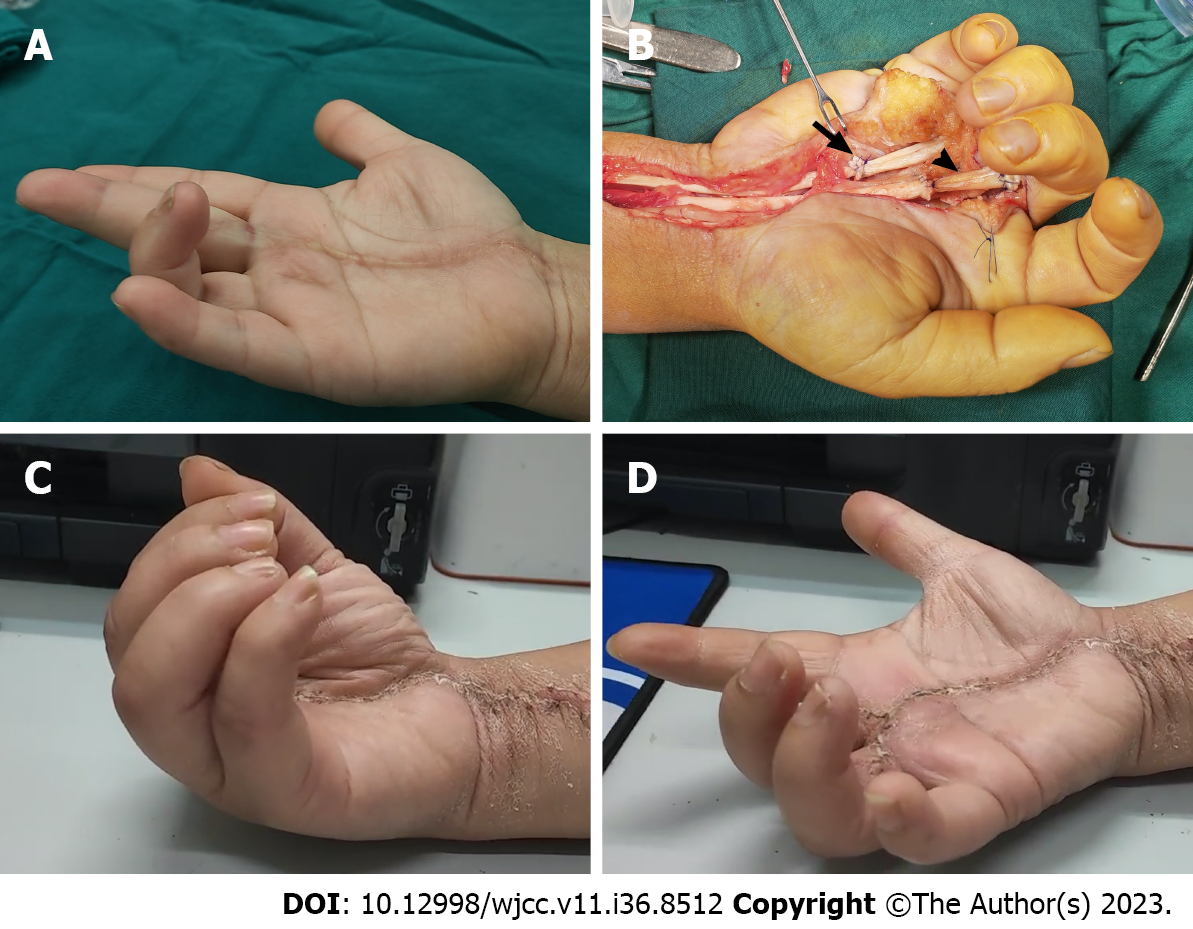
Figure 5 The second surgery and follow-up.
A: The extended middle finger and flexed ring finger before reexploration; B: Intraoperative view of the repaired tendons of the middle and ring fingers. Arrow: The repaired site of the ring finger with suturing the distal end of the flexor digitorum profundus to the proximal end of the flexor digitorum superficialis. Arrowhead: The repaired site of the middle finger with tendon grafting; C and D: Range of active motion of the affected fingers 10 mo after reoperation.
- Citation: Yan R, Zhang Z, Wu L, Wu ZP, Yan HD. Iatrogenic flexor tendon rupture caused by misdiagnosing sarcoidosis-related flexor tendon contracture as tenosynovitis: A case report. World J Clin Cases 2023; 11(36): 8512-8518
- URL: https://www.wjgnet.com/2307-8960/full/v11/i36/8512.htm
- DOI: https://dx.doi.org/10.12998/wjcc.v11.i36.8512


