Copyright
©The Author(s) 2023.
World J Clin Cases. Dec 26, 2023; 11(36): 8507-8511
Published online Dec 26, 2023. doi: 10.12998/wjcc.v11.i36.8507
Published online Dec 26, 2023. doi: 10.12998/wjcc.v11.i36.8507
Figure 1 Computed tomography.
A and B: Computed tomography examination of the thoracic spine showed bone destruction of the T4-5 vertebral body, the right pedicle and lamina of T5; C-E: Cervical, thoracic and lumbar magnetic resonance imaging showed multiple nodules with high signals on fat-suppressed images in the paravertebral and subcutaneous regions; F and G: High signals in the right vertebral body of T4-5, the right pedicle and lamina of T5 and high signals in the vertebral canal.
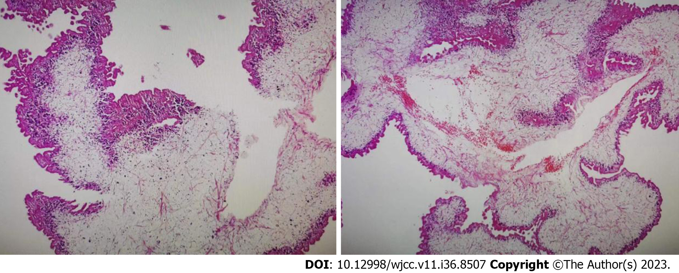
Figure 2 High power microscopic view of the parasite.
The body of the parasite has degenerated and calcareous bodies are not readily discerned, but the outer layer of the tegument was preserved.
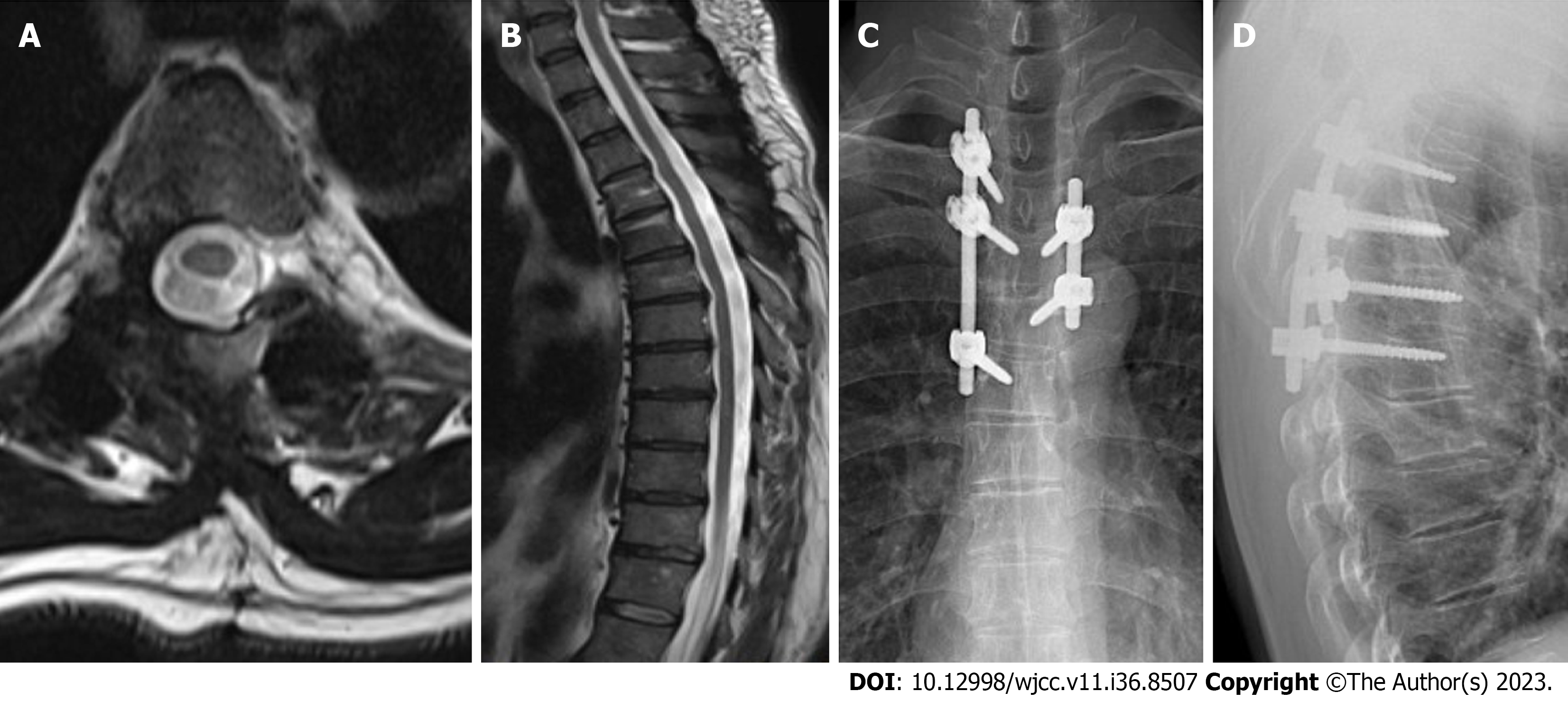
Figure 3 Magnetic resonance imaging.
A and B: At nine months after surgery, magnetic resonance imaging showed significant absorption of the lesion; C and D: Anteroposterior and lateral radiographs suggested that local curvature and the screw positions were satisfactory.
- Citation: Wen GJ, Chen J, Zhang SF, Zhou ZS, Jiao GL. Multiple sparganosis spinal infections mainly in the thoracic region: A case report. World J Clin Cases 2023; 11(36): 8507-8511
- URL: https://www.wjgnet.com/2307-8960/full/v11/i36/8507.htm
- DOI: https://dx.doi.org/10.12998/wjcc.v11.i36.8507


