Copyright
©The Author(s) 2023.
World J Clin Cases. Dec 26, 2023; 11(36): 8434-8439
Published online Dec 26, 2023. doi: 10.12998/wjcc.v11.i36.8434
Published online Dec 26, 2023. doi: 10.12998/wjcc.v11.i36.8434
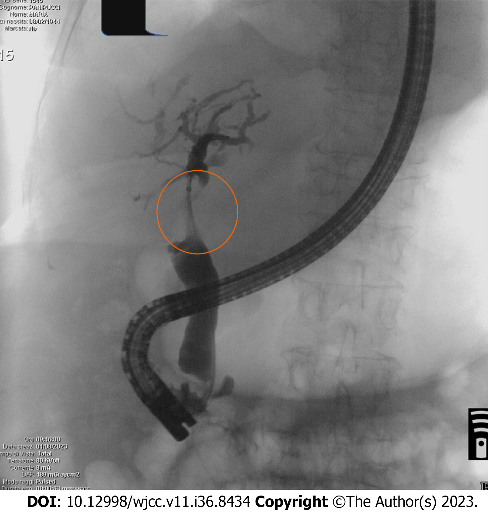
Figure 1 Endoscopic retrograde cholangiopancreatography performed at admission to clarify the radiological findings showed a serrated biliary hilar stenosis (orange circle) and dilatation of intrahepatic bile ducts.
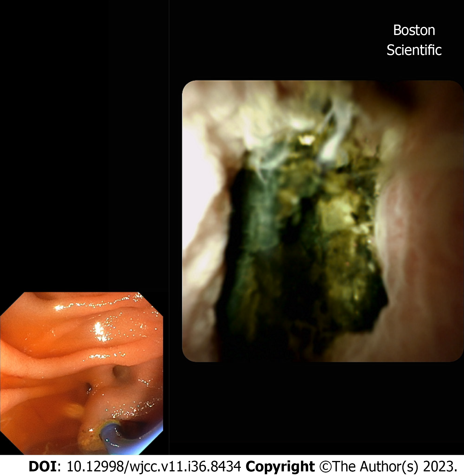
Figure 2 Choledoscopy with Spyglass II revealed the presence of fixed tissue in the hepatic hilum causing bile duct stenosis.
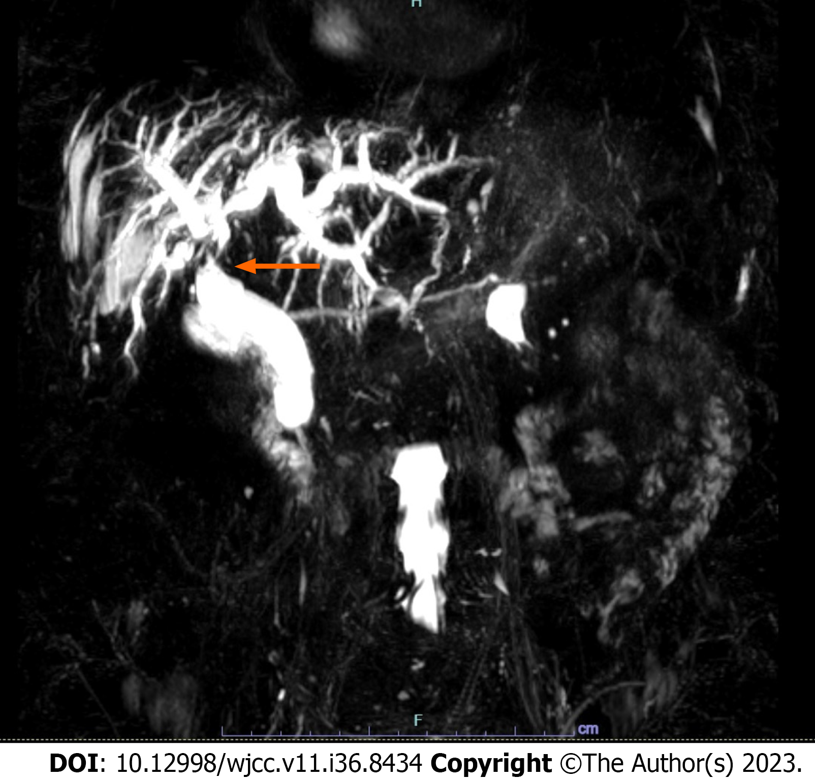
Figure 3 Magnetic resonance imaging cholangiopancreatography showing persistence of bile duct and intrahepatic biliary dilation after endoscopic treatment.
- Citation: Cocca S, Carloni L, Marocchi M, Grande G, Bianchini M, Colecchia A, Conigliaro R, Bertani H. Post-trans-arterial chemoembolization hepatic necrosis and biliary stenosis: Clinical charateristics and endoscopic approach. World J Clin Cases 2023; 11(36): 8434-8439
- URL: https://www.wjgnet.com/2307-8960/full/v11/i36/8434.htm
- DOI: https://dx.doi.org/10.12998/wjcc.v11.i36.8434


