Copyright
©The Author(s) 2023.
World J Clin Cases. Dec 16, 2023; 11(35): 8425-8430
Published online Dec 16, 2023. doi: 10.12998/wjcc.v11.i35.8425
Published online Dec 16, 2023. doi: 10.12998/wjcc.v11.i35.8425
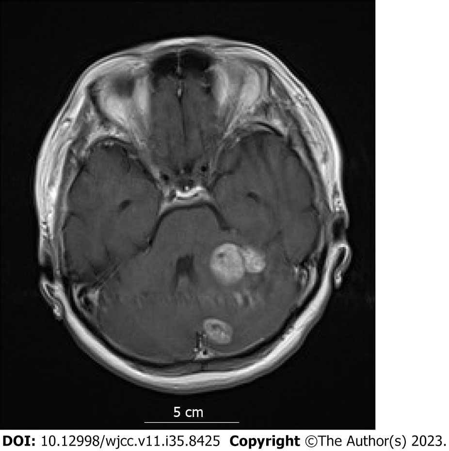
Figure 1 Initial head magnetic resonance imaging.
The patient was diagnosed with multiple brain metastases based on contrast-enhanced head magnetic resonance imaging when she visited our hospital for the staggering that she began experiencing in the 11th year postoperatively.
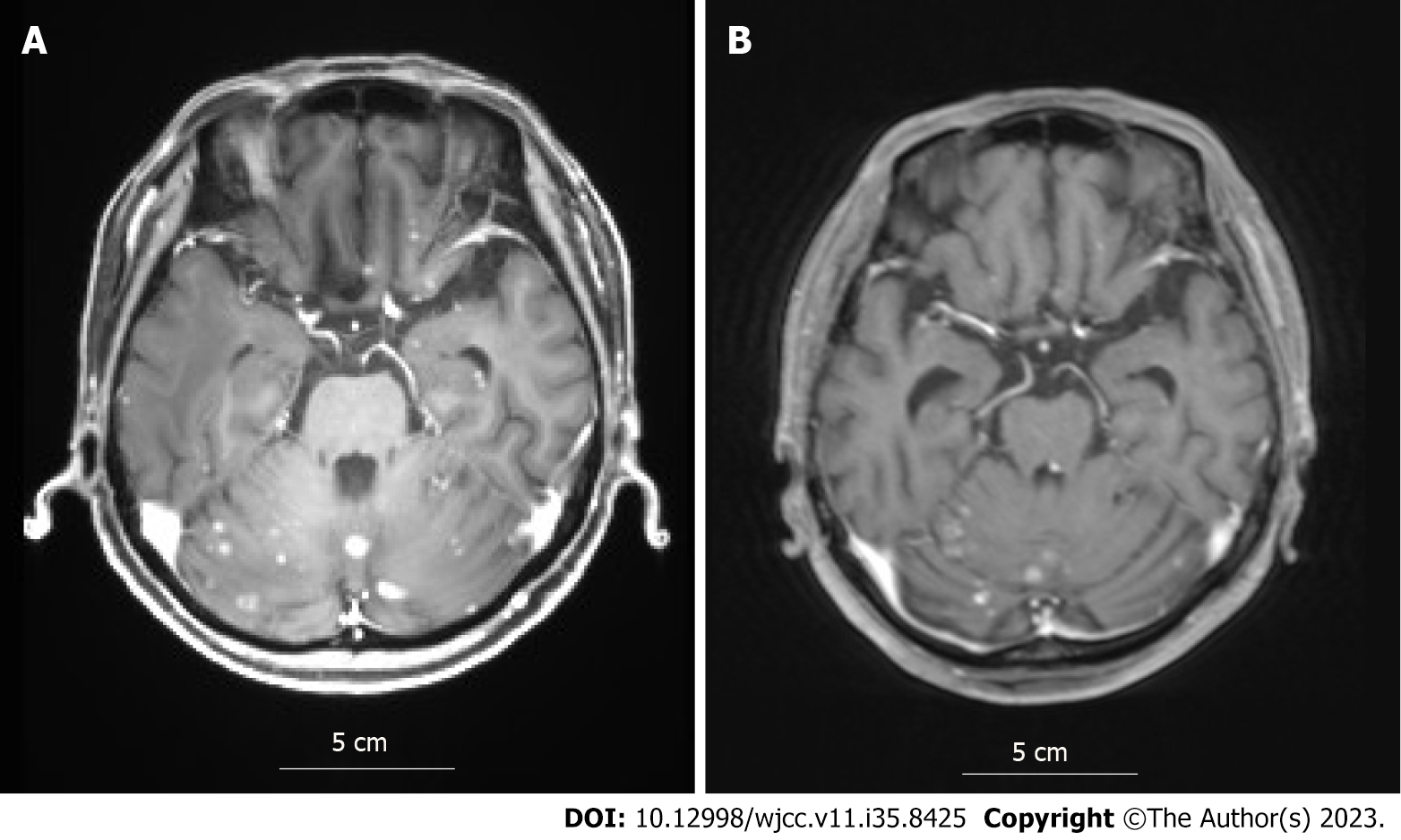
Figure 2 Magnetic resonance imaging before and after abemaciclib administration.
A: Contrast-enhanced head magnetic resonance imaging (MRI) before abemaciclib plus letrozole treatment; B: Contrast-enhanced head MRI after abemaciclib plus letrozole treatment. The brain metastases shrunk, and the contrast effect was attenuated after treatment, almost reaching a partial response.
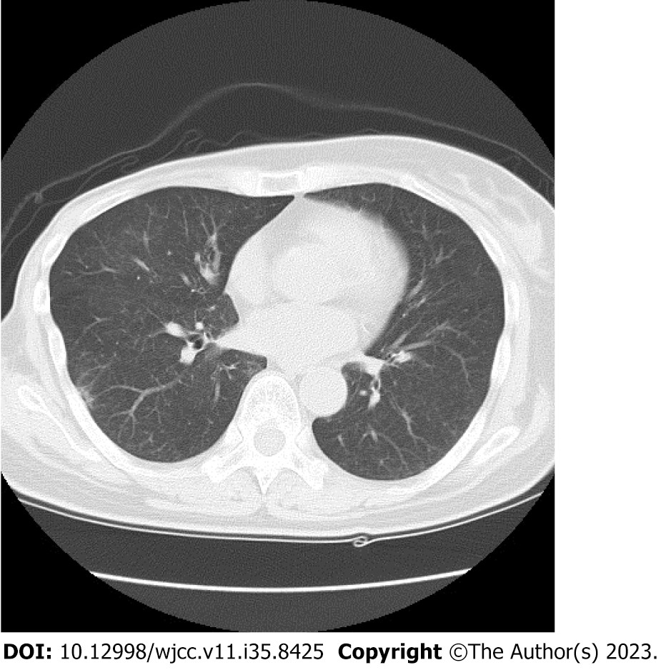
Figure 3 Computed tomography scan when diagnosing drug-induced lung disease.
Computed tomography after abemaciclib plus letrozole treatment reveals a pale interstitial shadow on the right lung, which led to the diagnosis of drug-induced lung disease.
- Citation: Yamashiro H, Morii N. Abemaciclib-induced lung damage leading to discontinuation in brain metastases from breast cancer: A case report. World J Clin Cases 2023; 11(35): 8425-8430
- URL: https://www.wjgnet.com/2307-8960/full/v11/i35/8425.htm
- DOI: https://dx.doi.org/10.12998/wjcc.v11.i35.8425


