Copyright
©The Author(s) 2023.
World J Clin Cases. Dec 6, 2023; 11(34): 8176-8183
Published online Dec 6, 2023. doi: 10.12998/wjcc.v11.i34.8176
Published online Dec 6, 2023. doi: 10.12998/wjcc.v11.i34.8176
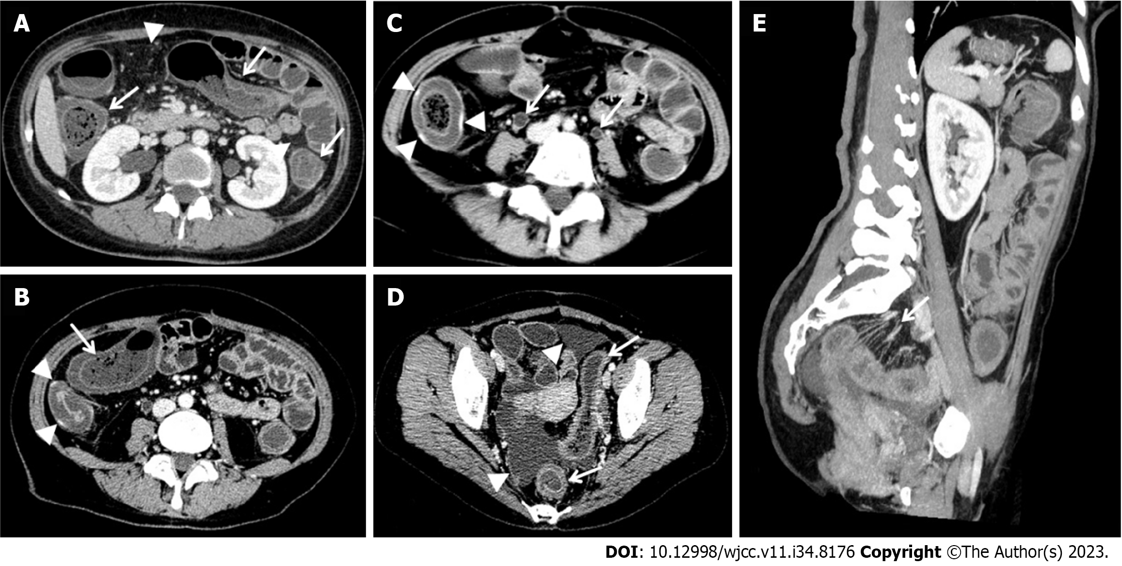
Figure 1 Contrast-enhanced computed tomography scan of our patient shows severe whole colon and rectum wall thickening, with the target sign or doughnut sign (white arrows).
A: The image shows the air-liquid level in the ascending colon, increased attenuation of mesenteric fat (white arrows head); B: The image shows the hyperemic ascending colon with obvious enhancement (white arrows head) and punctured gas breaking through the intestinal wall (white arrows); C: The image shows the ascending colon with ischemic and congestive changes in the same ascending bowel, which the hyperemic area showed obvious enhancement (white arrows head), and bilateral ureter-hydronephrosis (white arrows); D: The image shows rectum and sigmoid colon wall thickening with doughnut sign (white arrows), and ascites (white arrows head); E: The reconstructed coronal image shows mesenteric vessels enlargement with comb sign (white arrows).
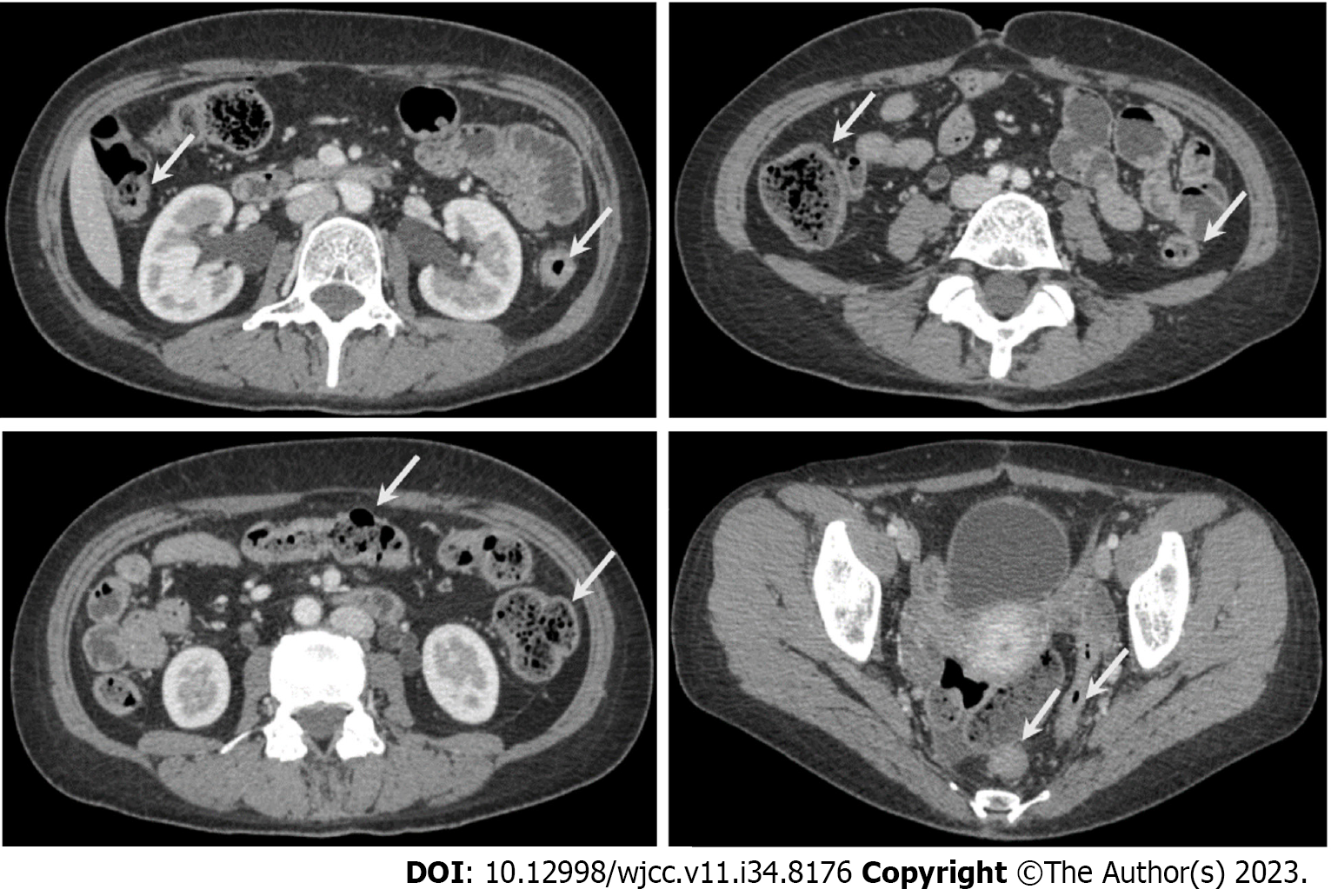
Figure 2 Contrast-enhanced computed tomography scan of our patient shows all colon is normal after one week of treatment (white arrows).
Figure 3 The treatment process for this case.
CT: Computed tomography.
- Citation: Gan H, Wang F, Gan Y, Wen L. Rare case of lupus enteritis presenting as colorectum involvement: A case report and review of literature. World J Clin Cases 2023; 11(34): 8176-8183
- URL: https://www.wjgnet.com/2307-8960/full/v11/i34/8176.htm
- DOI: https://dx.doi.org/10.12998/wjcc.v11.i34.8176


