Copyright
©The Author(s) 2023.
World J Clin Cases. Dec 6, 2023; 11(34): 8164-8169
Published online Dec 6, 2023. doi: 10.12998/wjcc.v11.i34.8164
Published online Dec 6, 2023. doi: 10.12998/wjcc.v11.i34.8164
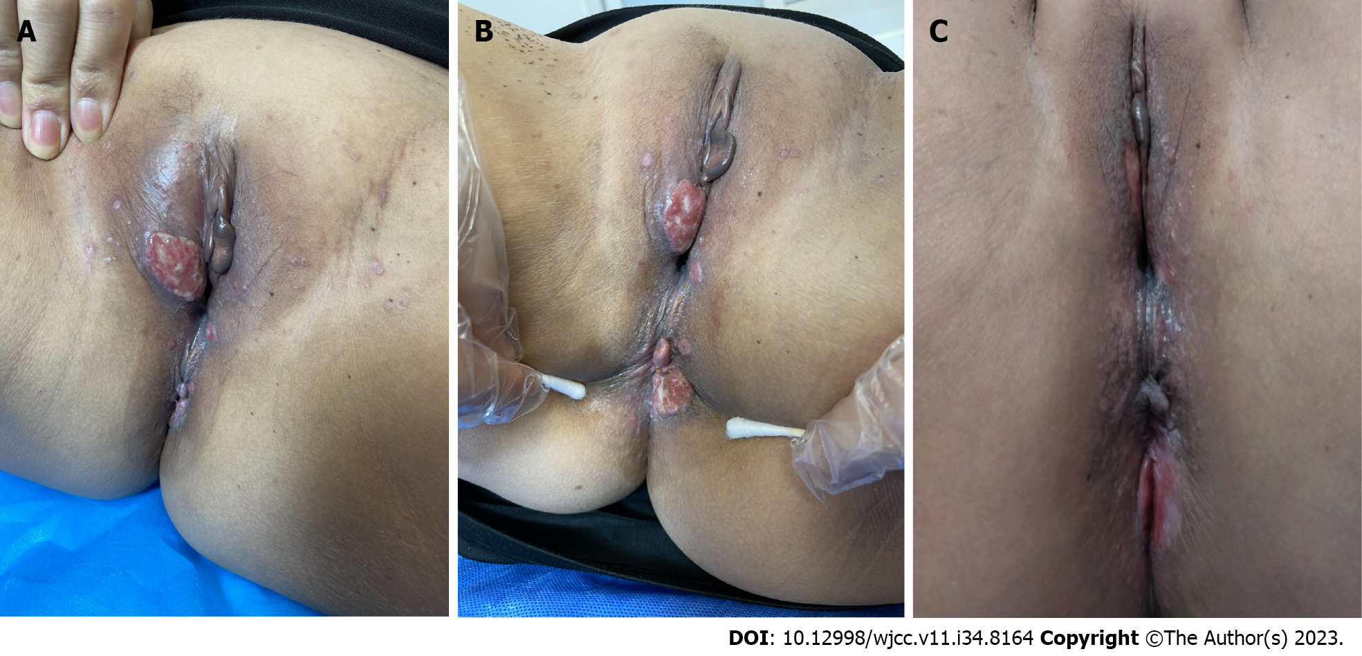
Figure 1 Clinical feature.
A: Red erosive plaques and ulcers on the vulva; B: Red erosive plaques and ulcers on the perianal area; C: Lesions after 6 mo of oral prednisone 20 mg orally.
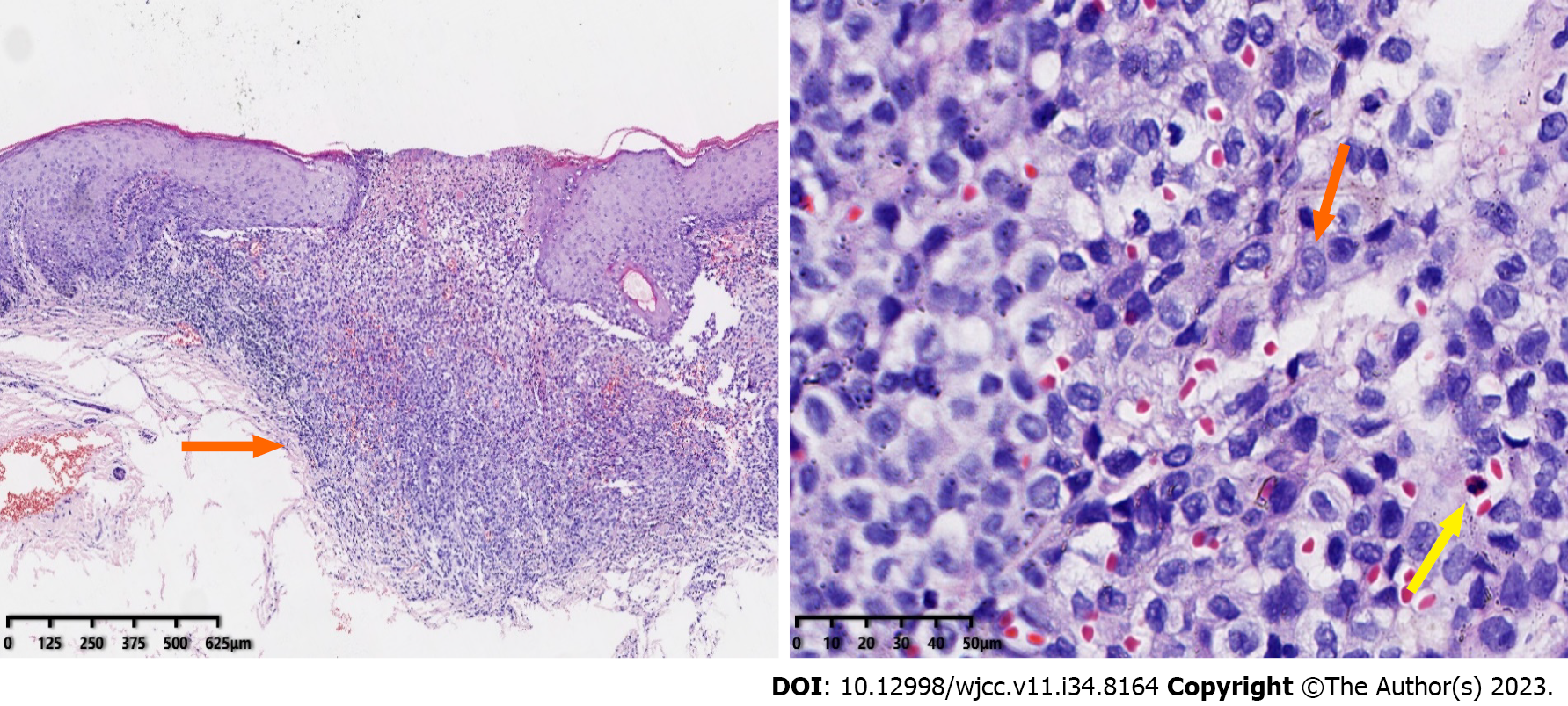
Figure 2 Hematoxylin and eosin staining.
A: The histiocytoid cells in the superficial and middle dermis layer with mild nuclear atypia, with occasional eosinophils; B: The histiocytoid cells in the superficial and middle dermis layer with mild nuclear atypia (orange arrow), with occasional eosinophils (yellow arrow).
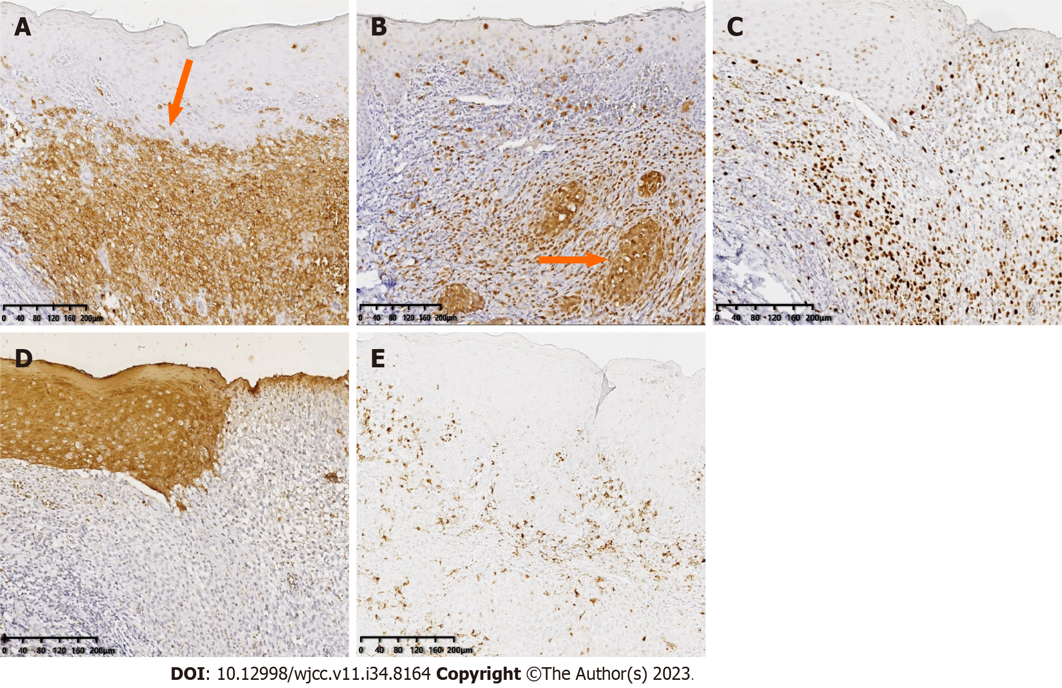
Figure 3 Immunohistochemistry.
A: CD1 (+++); B: S100 (+++); C: Ki-67 (30%); D: CK (-); E: CD68 (20%+).
- Citation: Yang PP, Hu SY, Chai XY, Shi XM, Liu LX, Li LE. Adult localized Langerhans cell histiocytosis: A case report. World J Clin Cases 2023; 11(34): 8164-8169
- URL: https://www.wjgnet.com/2307-8960/full/v11/i34/8164.htm
- DOI: https://dx.doi.org/10.12998/wjcc.v11.i34.8164


