Copyright
©The Author(s) 2023.
World J Clin Cases. Nov 26, 2023; 11(33): 8078-8083
Published online Nov 26, 2023. doi: 10.12998/wjcc.v11.i33.8078
Published online Nov 26, 2023. doi: 10.12998/wjcc.v11.i33.8078
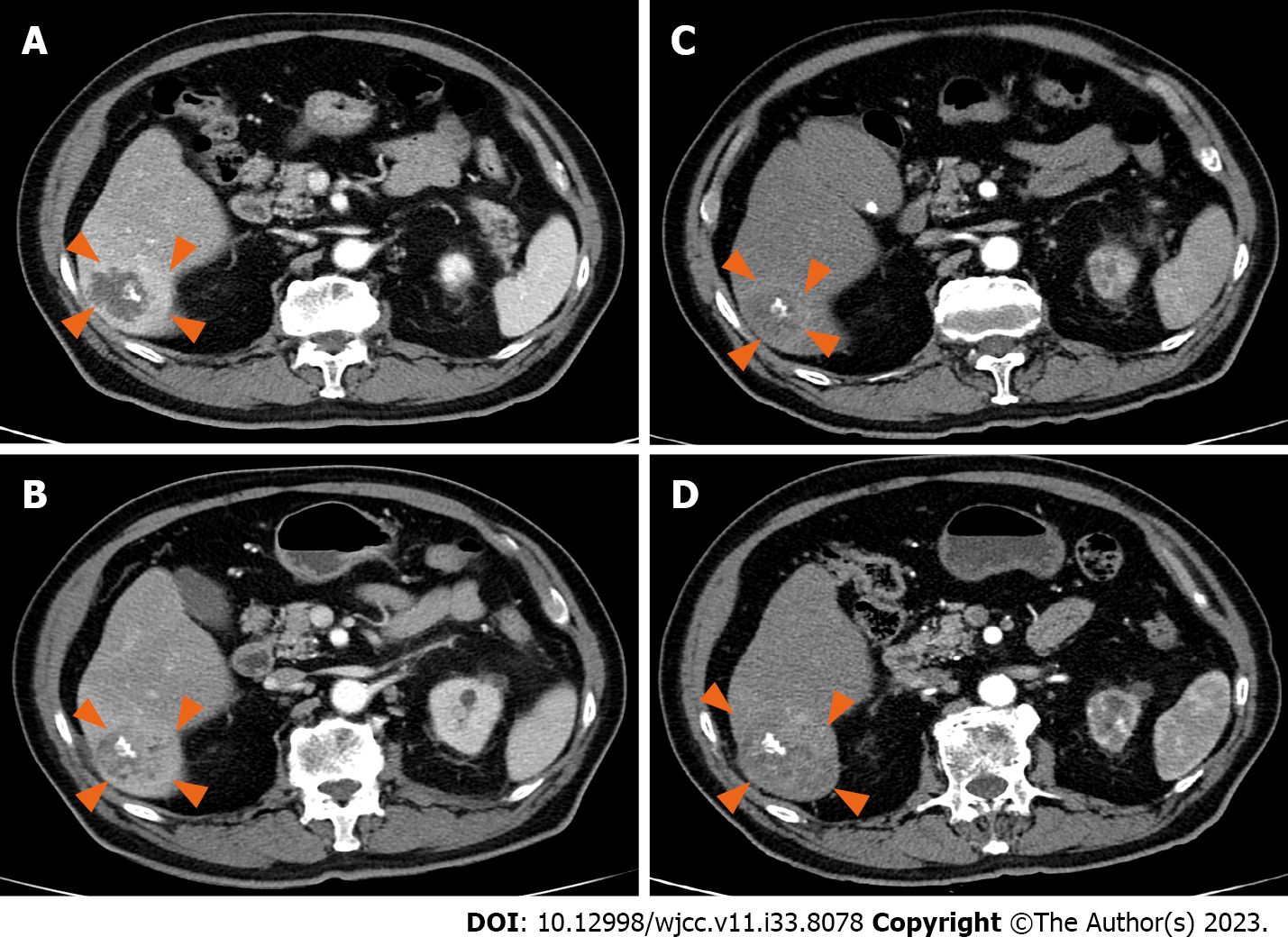
Figure 1 Liver computed tomography findings.
A: On admission day. A lobulated margined multiseptated with central septal calcified 3.9 cm cystic attenuating mass with surrounding prolonged hyperemia in hepatic segment VI; B: Computed tomography (CT) 1 mo later. Still noted was a lobulated margined septated and central calcified cystic attenuating lesion with surrounding high enhancement. The size decreased to 3.3 cm; C: CT at 6 mo. Slightly increased size (from 3.3 cm to 3.6 cm) of the lobulated margined delayed minimal heterogeneous enhancing lesion with surrounding hyperemia, without any interval change in the irregular margined focal central calcification; D: CT at 10 mo. The size of the mass markedly increased to 6 cm.
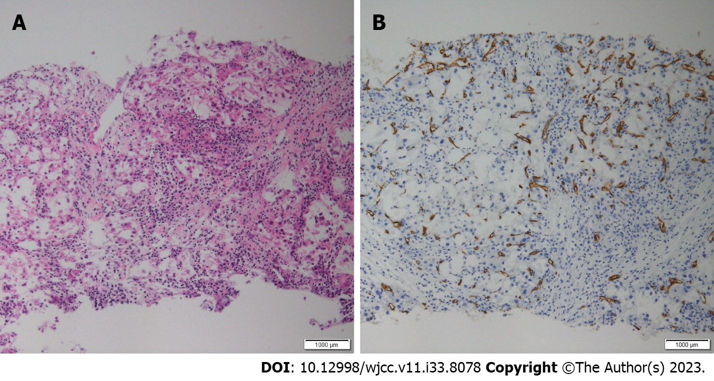
Figure 2 Liver biopsy histopathological findings.
A and B: On H&E staining (A), inflammatory cells and focal carcinomatous changes are observed; some areas show positive staining for CD34 (B).
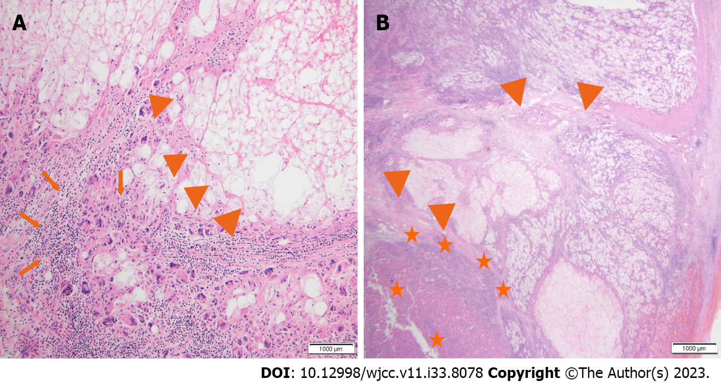
Figure 3 Postoperative histopathological findings.
A and B: A significant portion of the liver parenchyma has undergone necrosis, showing abscess-like features (arrowhead), inflammatory cell infiltration (arrow) and hepatocellular carcinoma tumor cells (star) in the remaining liver parenchyma.
- Citation: Ryou SH, Shin HD, Kim SB. Hepatocellular carcinoma presenting as organized liver abscess: A case report. World J Clin Cases 2023; 11(33): 8078-8083
- URL: https://www.wjgnet.com/2307-8960/full/v11/i33/8078.htm
- DOI: https://dx.doi.org/10.12998/wjcc.v11.i33.8078


