Copyright
©The Author(s) 2023.
World J Clin Cases. Nov 26, 2023; 11(33): 8030-8037
Published online Nov 26, 2023. doi: 10.12998/wjcc.v11.i33.8030
Published online Nov 26, 2023. doi: 10.12998/wjcc.v11.i33.8030
Figure 1 Abdominal contrast-enhanced computed tomography images.
Images show a huge cystic mass in the abdominal and pelvic cavities, possibly arising from the liver. A small amount of free fluid is present in the pelvic cavity. A: Middle abdomen; B: Lower abdomen; C: Pelvic cavity; D: Coronal plane of the abdomen; E: Sagittal plane of the abdomen.
Figure 2 Magnetic resonance imaging.
Images show a large cystic mass in the abdominal and pelvic cavities that was considered a benign lesion. A small amount of free fluid is present in the pelvic cavity. A: Pelvic cavity; B: Enhanced pelvic cavity; C: Sagittal plane of the abdomen; D: Enhanced sagittal plane of the abdomen.
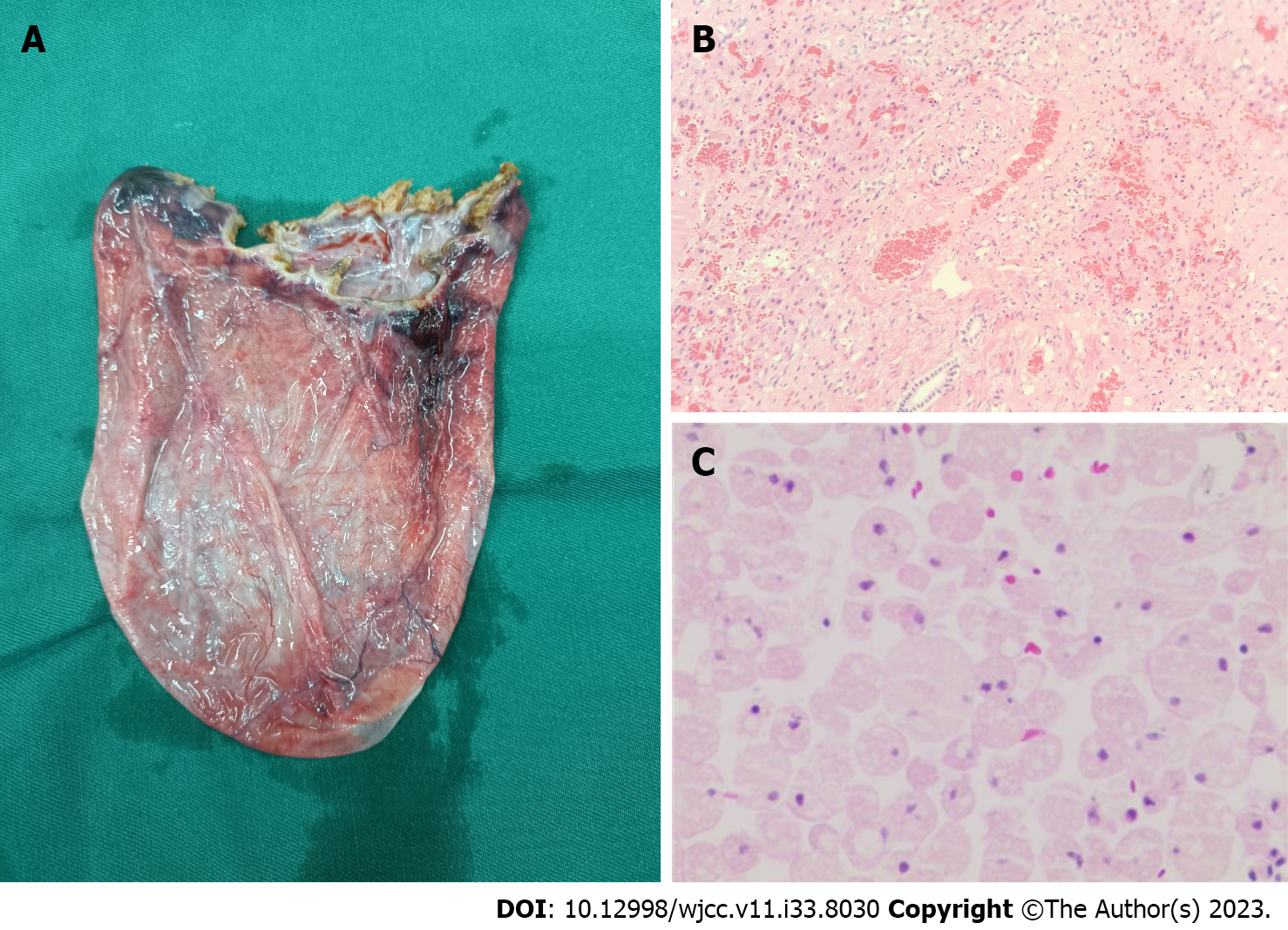
Figure 3 Postoperative specimen and pathological diagnosis.
A: Postoperative specimen; B: Bile duct derived cyst liver tissue showing bleeding and a compression injury (liver tissue); C: A few red blood cells and histiocytes (ascites cell mass).
- Citation: Li S, Tang J, Ni DS, Xia AD, Chen GL. Giant complex hepatic cyst causing pseudocystitis: A case report. World J Clin Cases 2023; 11(33): 8030-8037
- URL: https://www.wjgnet.com/2307-8960/full/v11/i33/8030.htm
- DOI: https://dx.doi.org/10.12998/wjcc.v11.i33.8030


