Copyright
©The Author(s) 2023.
World J Clin Cases. Nov 26, 2023; 11(33): 8022-8029
Published online Nov 26, 2023. doi: 10.12998/wjcc.v11.i33.8022
Published online Nov 26, 2023. doi: 10.12998/wjcc.v11.i33.8022
Figure 1 Computed tomography examination before surgery.
A: A ground-glass nodule with rough edges (about 2.5 mm × 9 mm in size) in the apicoposterior segment of left upper lobe; B: A ground-glass nodule with clear edges (about 6 mm × 4 mm in size) in the lateral basal segment of left lower lobe; C: Ground-glass density shadows were seen in the apex of right lungs, with blurred edges, and the range was approximately 9 mm × 6 mm.
Figure 2 High-resolution computed tomography 21 mo after surgery.
A: A pure ground-glass nodule in the apical segment of the right upper lobe (about 13.2 mm × 5.6 mm in size); B: A mixed ground glass in the superior segment of the right lower lobe Nodules (about 4.3 mm × 2.9 mm in size); C: A pure ground-glass nodule in the lateral basal segment of the left lower lobe (about 7.2 mm × 5.3 mm in size).
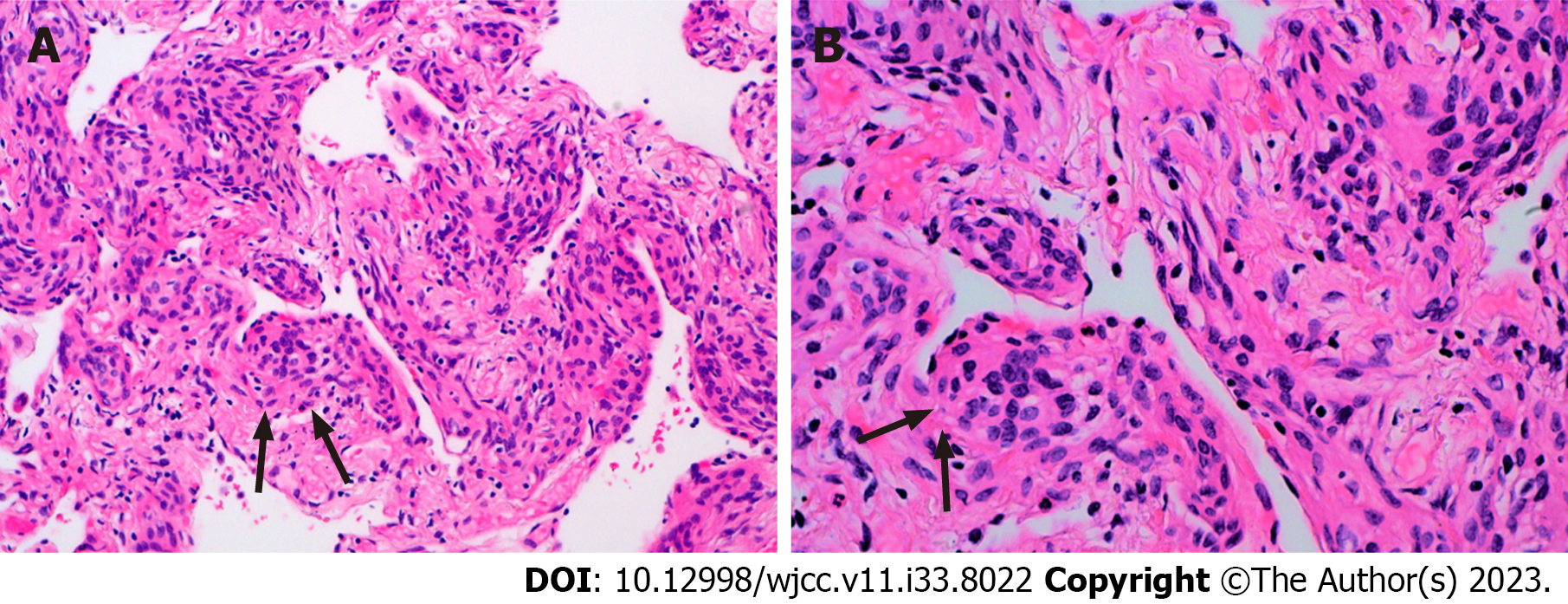
Figure 3 Histomorphological manifestation of right lower lobe nodule was the small foci in the alveolar septum were distributed in mild form of the aggregation of short spindle cells.
A: Original magnification 200×; B: Original magnification 400×.
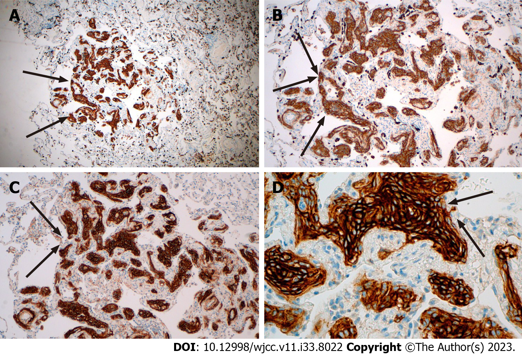
Figure 4 The Immunohistochemistry showed that the lesion was positive for epithelial membrane antigen and somatostatin receptor 2a.
A: The lesion was positive for epithelial membrane antigen, original magnification 200×; B: The lesion was positive for epithelial membrane antigen, original magnification 400×; C: The lesion was positive for somatostatin receptor 2a, original magnification 200×; D: The lesion was positive for somatostatin receptor 2a, original magnification 400×.
- Citation: Ruan X, Wu LS, Fan ZY, Liu Q, Yan J, Li XQ. Pathological diagnosis and immunohistochemical analysis of minute pulmonary meningothelial-like nodules: A case report. World J Clin Cases 2023; 11(33): 8022-8029
- URL: https://www.wjgnet.com/2307-8960/full/v11/i33/8022.htm
- DOI: https://dx.doi.org/10.12998/wjcc.v11.i33.8022


