Copyright
©The Author(s) 2023.
World J Clin Cases. Nov 16, 2023; 11(32): 7926-7934
Published online Nov 16, 2023. doi: 10.12998/wjcc.v11.i32.7926
Published online Nov 16, 2023. doi: 10.12998/wjcc.v11.i32.7926
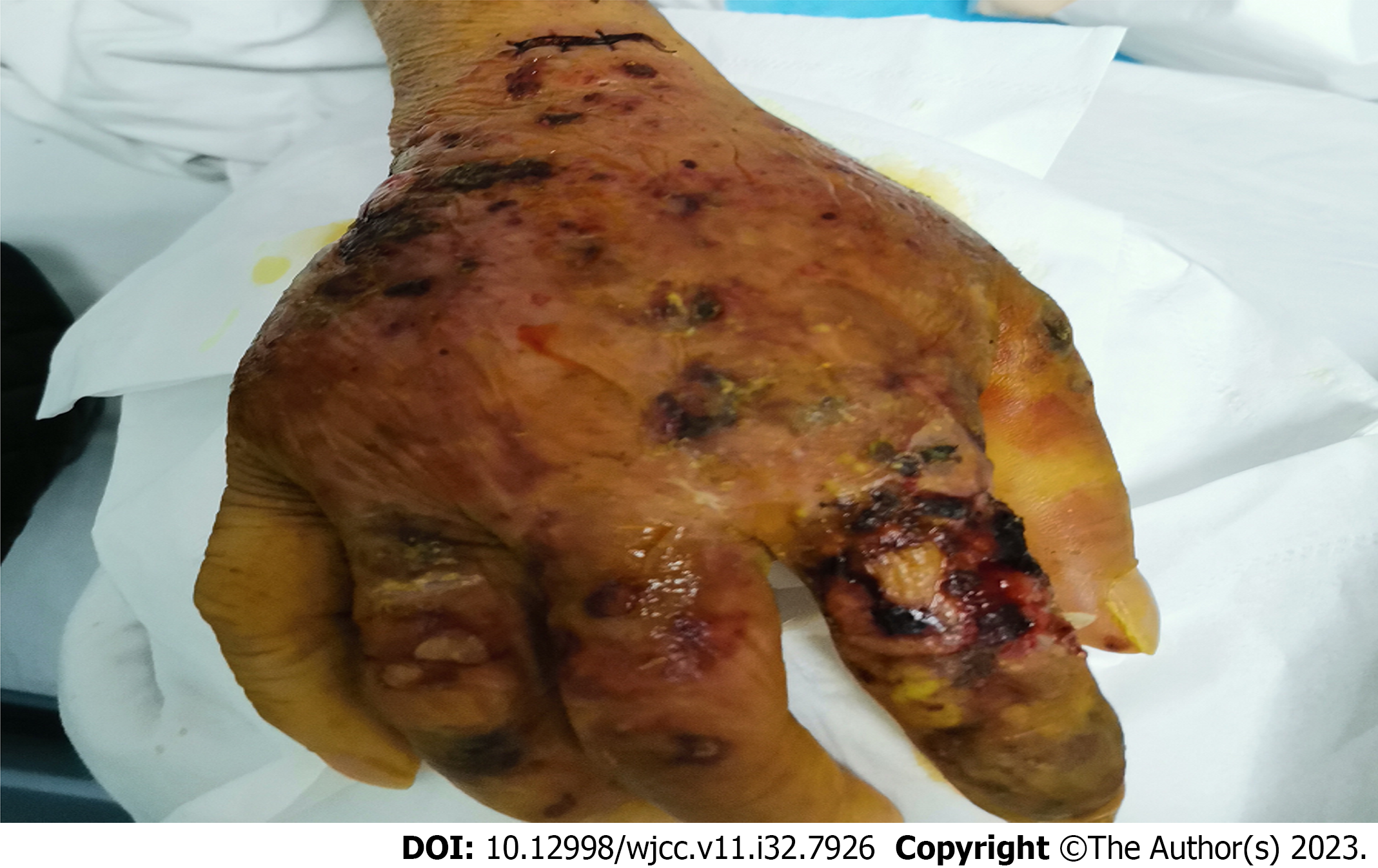
Figure 1 Skin lesions on the patient's right hand.
The rash appeared on the right upper limb, with partial blisters and ulceration, accompanied by pus flow and blackness.
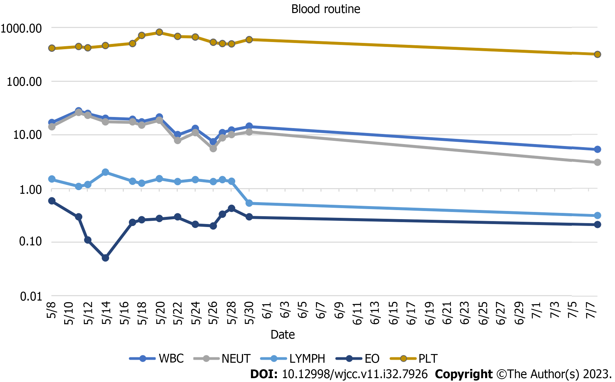
Figure 2 The changes in the blood routine indexes of the patient.
The patient's white blood cells and neutrophils were significantly elevated at the time of admission and they gradually decreased to normal after treatment. Eosinophils and lymphocytes had only a little effect during the treatment course, while the platelets were first elevated and then decreased to the normal counts. WBC: White blood cells; NEUT: Neutrophils; LYMPH: Lymphocyte; EO: Eosinophil; PLT: Platelet.
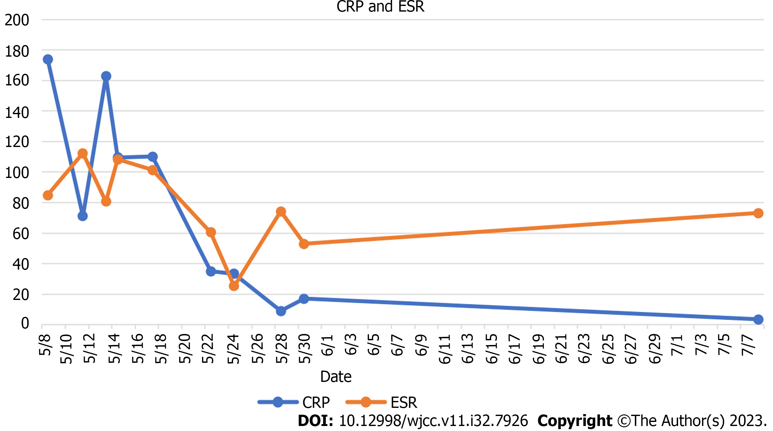
Figure 3 C-reactive protein and erythrocyte sedimentation rate of the patients.
After treatment, the levels of C-reactive protein (CRP) and erythrocyte sedimentation rate (ESR) decreased significantly when compared to that at the time of admission (CRP normal range 0-3 mg/L, ESR normal range 0-20 mm/h). CRP: C-reactive protein, ESR: Erythrocyte sedimentation rate.
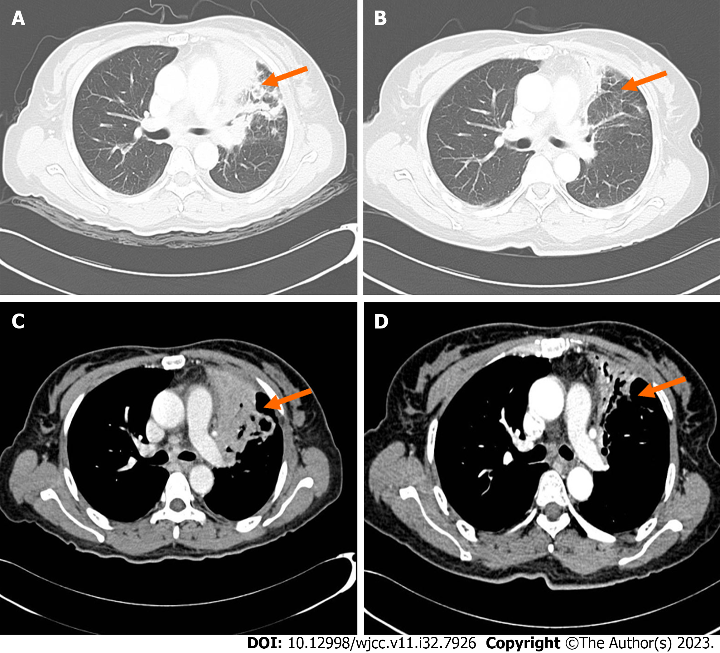
Figure 4 Chest computed tomography enhancement.
A: Lung window of 2021-05-10 (orange arrow); B: Lung window of 2021-07-12 (orange arrow); C: Mediastinal fenestra of 2021-05-10 (orange arrow); D: Mediastinal fenestra of 2021-07-12 2021-05-10 chest computed tomography (CT) revealed a high-density mass of size 7.7 cm × 7.2 cm × 5.9 cm in the upper lobe of the left lung in the lung window and lymph node enlargement in the mediastinal window. 2021-07-12 chest CT indicates improvement in the pulmonary lesion after treatment (orange arrow).
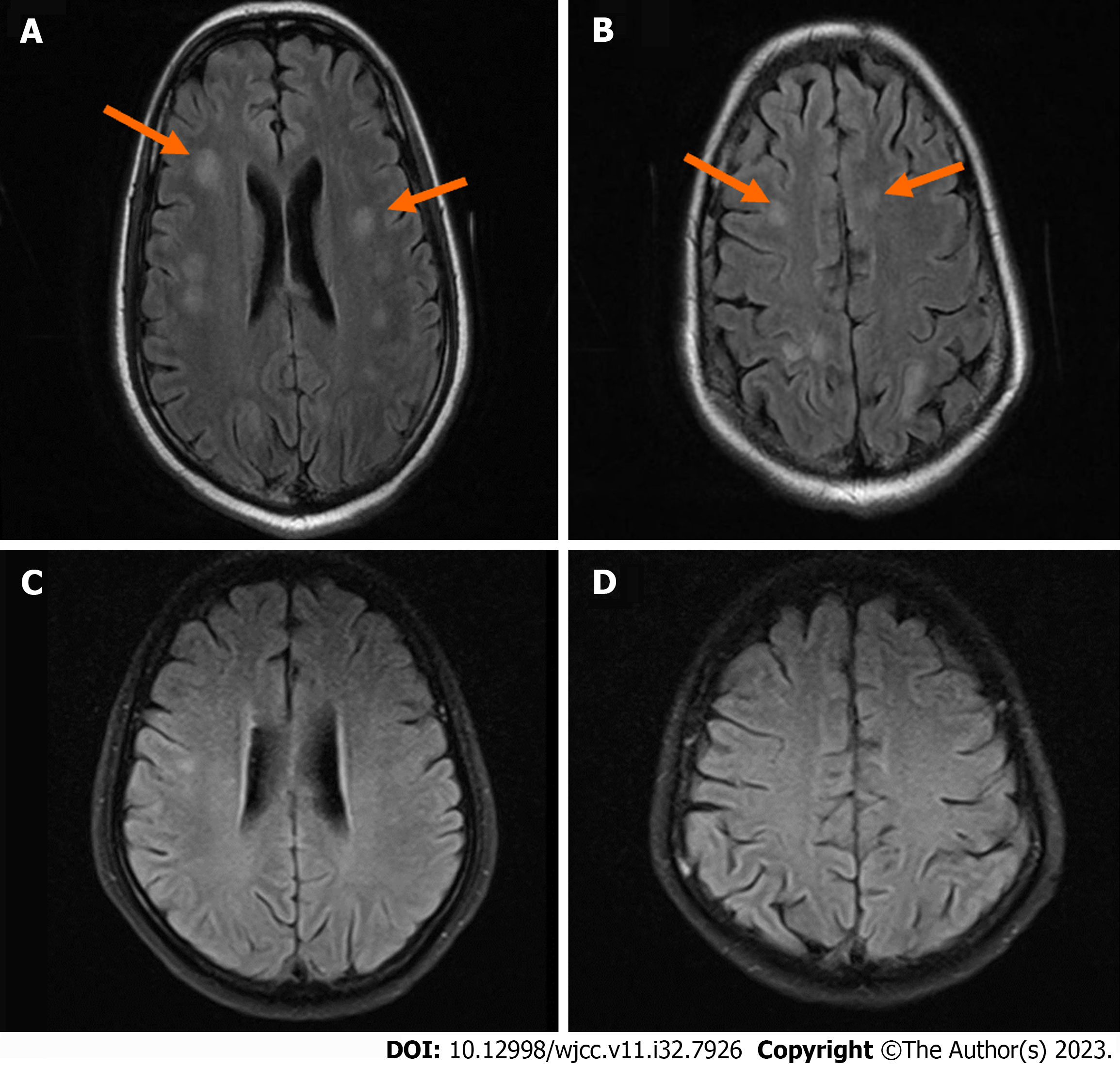
Figure 5 Brain magnetic resonance imaging.
A and B: Brain magnetic resonance imaging (MRI) of 2021-05-11 (orange arrows); C and D: Brain MRI of 2021-07-09 2021-05-11 brain MRI scan plus: T2 flair enhancement revealed multiple ischemic lesions in the right basal ganglia, bilateral frontal lobes, peri-ventricular, radiative crown, and hemioval center. 2021-07-09 brain MRI indicates improvement in the brain after treatment.
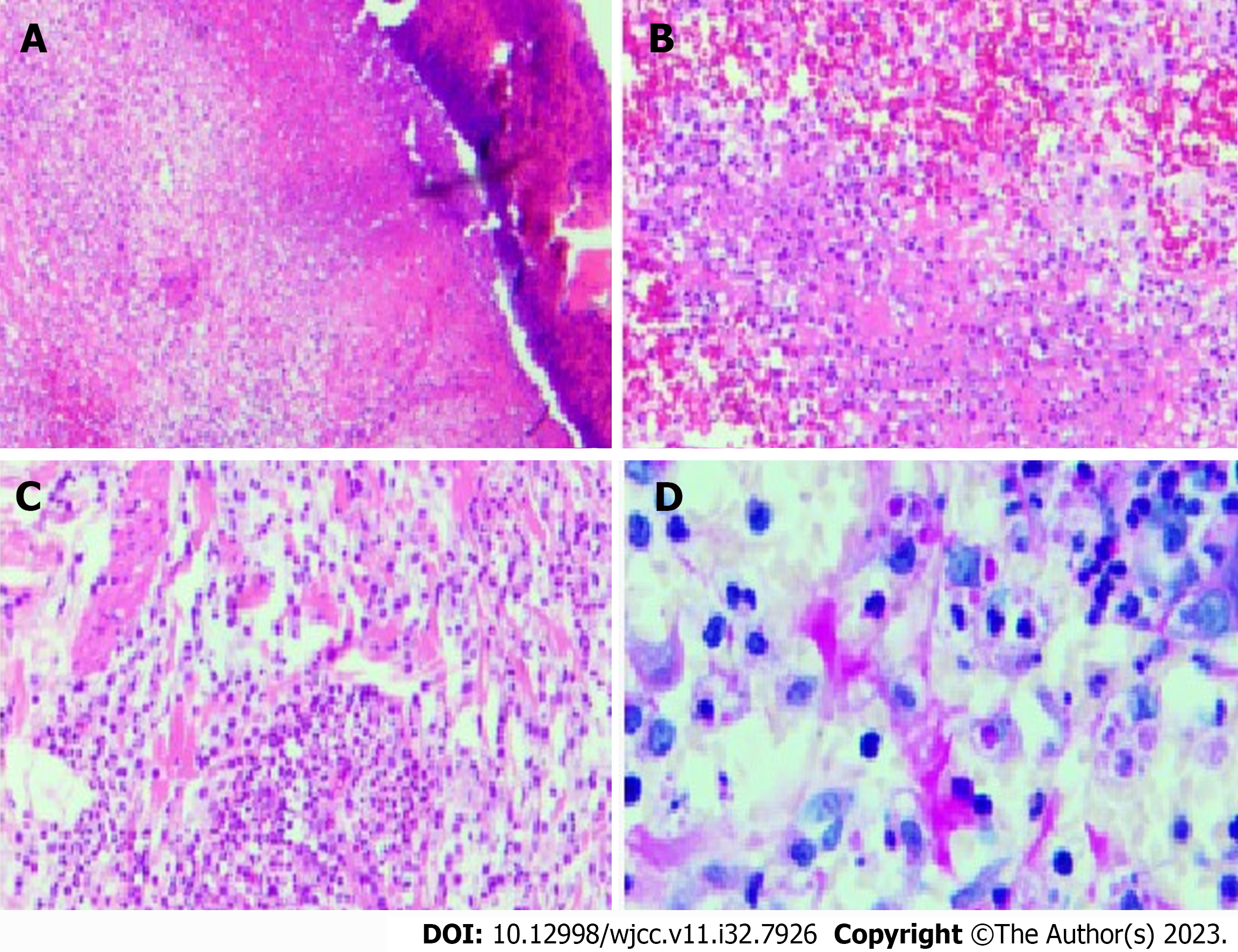
Figure 6 Hepatic encephalopathy staining of the right-hand lesion skin.
A: × 5; B: × 10; C: × 20; D: × 40. Epidermal erosion was observed by microscopy. Multiple focal necrosis were observed in the dermis, accompanied by further infiltration of lymphocytes and neutrophils. No epithelioid cells and caseous necrosis were observed. The observation was consistent with that of skin infection.
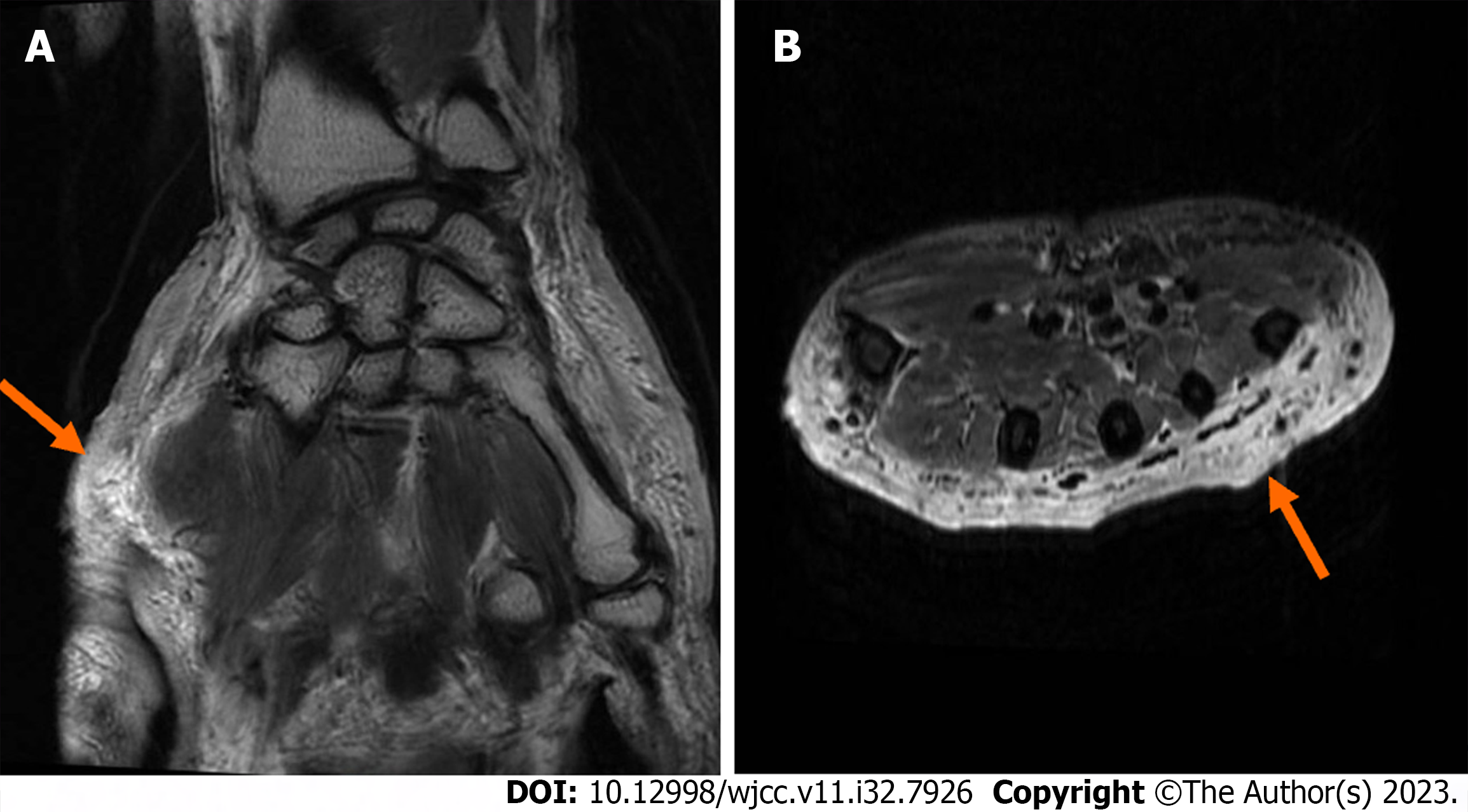
Figure 7 Right-hand magnetic resonance imaging.
A: Coronal position of the right hand magnetic resonance imaging (MRI) of 2021-5-15 (orange arrow); B: Axial position of the right hand MRI of 2021-5-15 2021-05-15 right-hand MRI indicates a slight swelling of the right hand; the subcutaneous fat space of the right palm was slightly blurred, implying the possibility of inflammatory changes (orange arrow).
- Citation: Zheng JH, Wu D, Guo XY. Intracranial infection accompanied sweet’s syndrome in a patient with anti-interferon-γ autoantibodies: A case report. World J Clin Cases 2023; 11(32): 7926-7934
- URL: https://www.wjgnet.com/2307-8960/full/v11/i32/7926.htm
- DOI: https://dx.doi.org/10.12998/wjcc.v11.i32.7926


