Copyright
©The Author(s) 2023.
World J Clin Cases. Nov 16, 2023; 11(32): 7852-7857
Published online Nov 16, 2023. doi: 10.12998/wjcc.v11.i32.7852
Published online Nov 16, 2023. doi: 10.12998/wjcc.v11.i32.7852
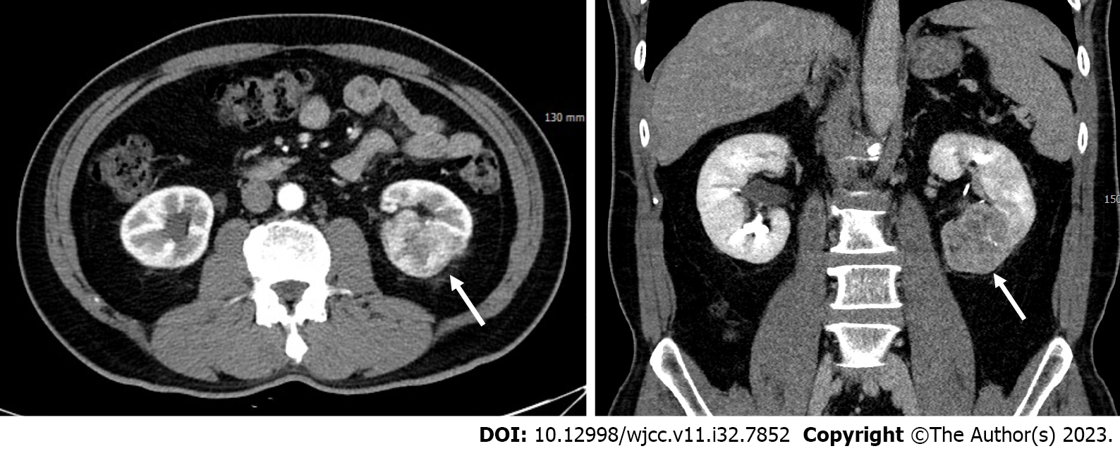
Figure 1 Pre-operative computed tomography.
Pre-operative computed tomography images demonstrating a heterogeneously enhancing mass (arrows) located in the lower polar area of the left kidney, suggestive of renal cell carcinoma.
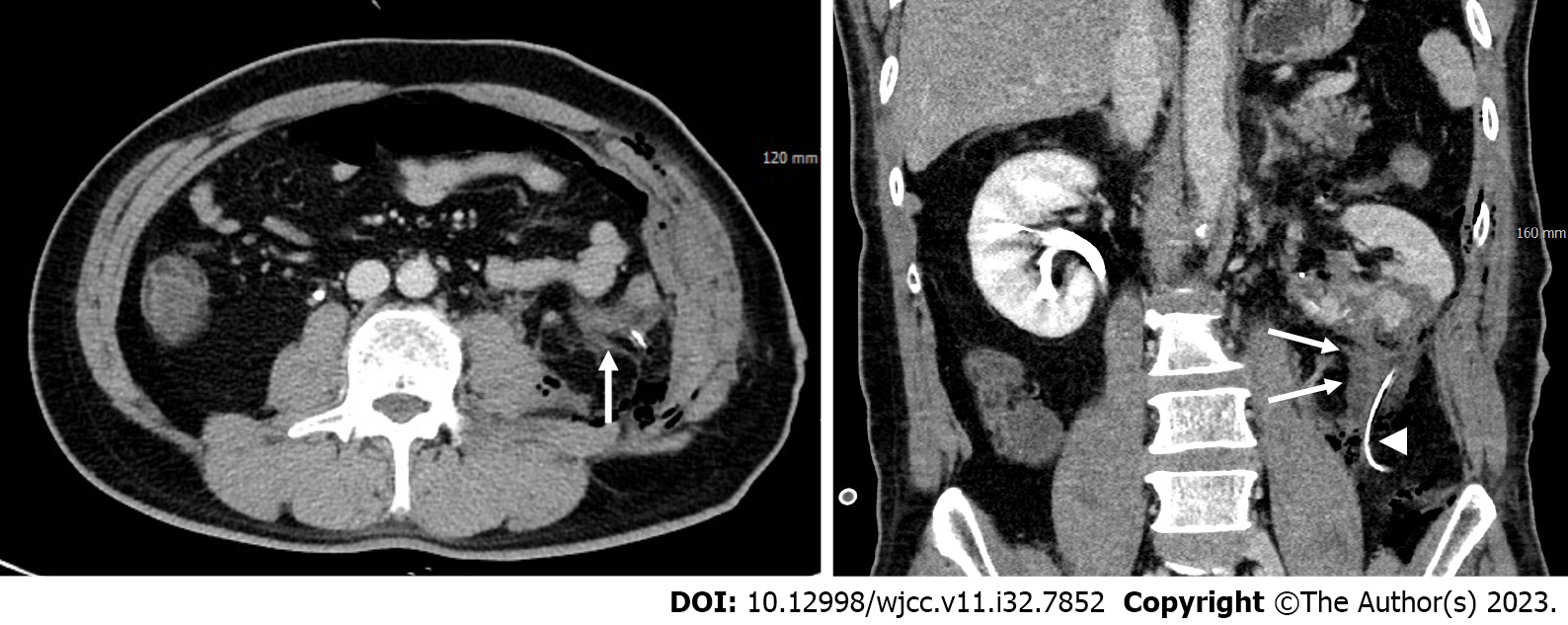
Figure 2 Post-operative computed tomography.
Post-operative computed tomography images obtained 1 d after partial nephrectomy revealing a small amount of fluid (arrows) in the inferior aspect of the left kidney, adjacent to the drainage tube (arrowhead), without evidence of contrast extravasation.
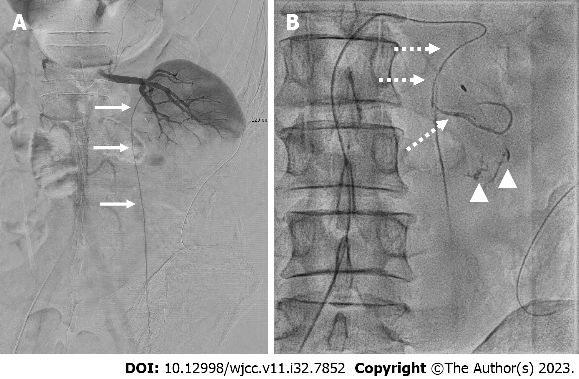
Figure 3 Transcatheter angiography.
A: Digital subtraction angiography of the left renal artery demonstrating no evidence of active bleeding and revealing the left testicular artery (arrows) arising from the middle segmental artery of the renal artery; B: Fluoroscopic spot image obtained following super-selective catheterization (dashed arrows) of the suspected branch arising from the testicular artery, revealing contrast extravasation (arrowheads).
Figure 4 Post-embolization images.
A: Fluoroscopic spot image obtained after transcatheter embolization, demonstrating cast formation (arrows) of n-butyl-2-cyanoacrylate and iodized oil mixture at the bleeding site; B and C: Post-embolization digital subtraction angiography illustrating preserved distal flow of the renal artery (B) and testicular artery (C).
- Citation: Youm J, Choi MJ, Kim BM, Seo Y. Transcatheter embolization for hemorrhage from aberrant testicular artery after partial nephrectomy: A case report. World J Clin Cases 2023; 11(32): 7852-7857
- URL: https://www.wjgnet.com/2307-8960/full/v11/i32/7852.htm
- DOI: https://dx.doi.org/10.12998/wjcc.v11.i32.7852


