Copyright
©The Author(s) 2023.
World J Clin Cases. Nov 6, 2023; 11(31): 7706-7711
Published online Nov 6, 2023. doi: 10.12998/wjcc.v11.i31.7706
Published online Nov 6, 2023. doi: 10.12998/wjcc.v11.i31.7706
Figure 1 Endoscopic retrograde cholangiopancreatography examination and treatment.
A: Duodenal papilla hemorrhage revealed by endoscopic retrograde cholangiopancreatography; B: Placement of a biliary covered metal stent.
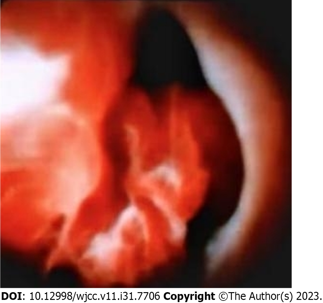
Figure 2 A neoplasm of the common bile duct with active bleeding was observed under a SpyGlass.
Figure 3 Malignant small round cell tumor with tumor cells showing some cytoplasm and difficult-to-see karyokinesis was observed under a light microscope following hematoxylin and eosin staining of the tumor tissue.
Figure 4 The SS18 gene break test by fluorescence in situ hybridization revealed mostly red and green markers in a fusion state and only a few red and green markers in a nonfusion state (fluorescence in situ hybridization-positive cells), indicating that the SS18 gene break test result was negative.
- Citation: Jin YL, Ruan YJ, Lu GR. Biliary hemorrhage caused by a malignant small round cell tumor in the common bile duct: A case report. World J Clin Cases 2023; 11(31): 7706-7711
- URL: https://www.wjgnet.com/2307-8960/full/v11/i31/7706.htm
- DOI: https://dx.doi.org/10.12998/wjcc.v11.i31.7706


