Copyright
©The Author(s) 2023.
World J Clin Cases. Nov 6, 2023; 11(31): 7699-7705
Published online Nov 6, 2023. doi: 10.12998/wjcc.v11.i31.7699
Published online Nov 6, 2023. doi: 10.12998/wjcc.v11.i31.7699
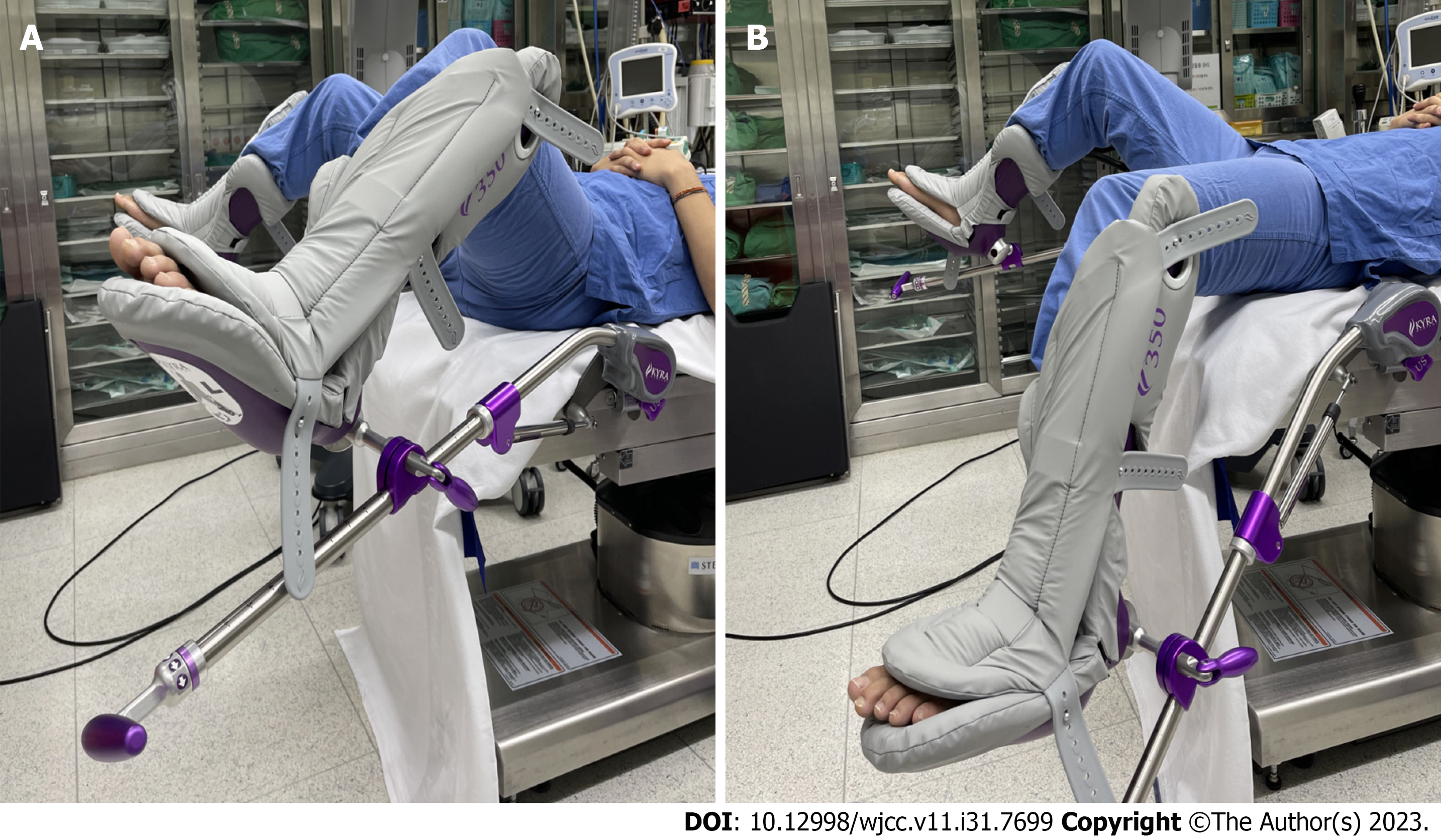
Figure 1 The positions adopted by the patient during the operation.
A: The lithotomy position with hip flexion 45°, knee flexion 45°, and thighs apart by 90°. The legs were fixed with support from the boots; B: The patient intermittently performed hip extension on the left side, extending the hip to the neutral position. The patient was maintained in the lithotomy position during surgery, but switched to a position where she extended her hip and leg on the left side several times, always returning to the lithotomy position thereafter. This repetitive posture change may have caused stretching and angulation of the lateral femoral cutaneous nerve.
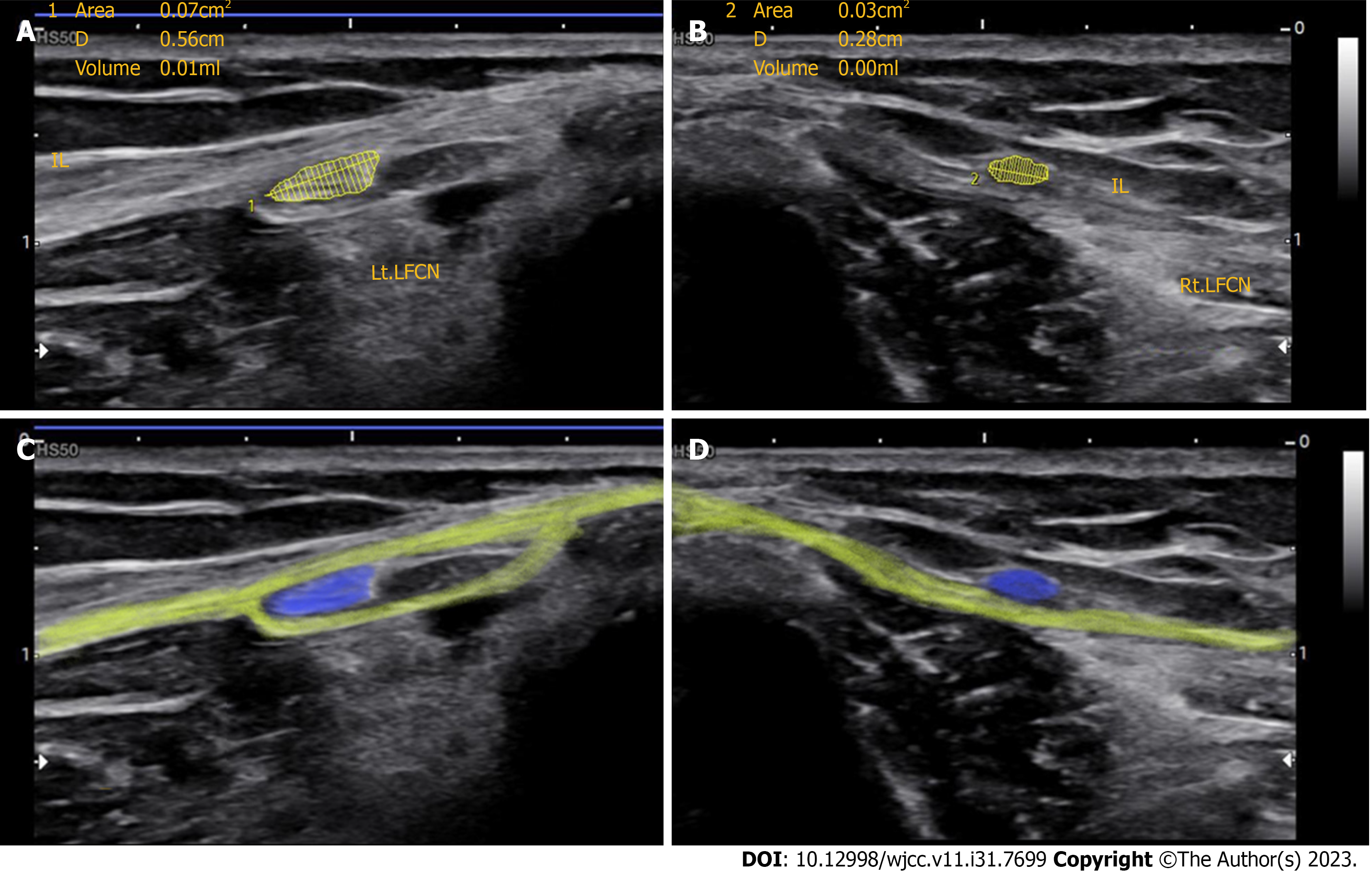
Figure 2 Left and right lateral femoral cutaneous nerves.
A: The left (Lt) lateral femoral cutaneous nerve (LFCN) passes through the inguinal ligament (IL); the nerve was swollen at the point of passage. The cross-sectional area (CSA) was 7 mm2, and the longitudinal diameter (LD) was 5.6 mm; B: The right (Rt) LFCN passes over the IL. The CSA was 3 mm2, and the LD was 2.8 mm; C: The blue shadow indicates the Lt IL, and the yellow shadow indicates the Lt LFCN. The LFCN passes through the IL; D: The blue shadow represents the Rt IL, and the yellow shadow indicates the Rt LFCN. The LFCN passes over the IL. IL: Inguinal ligament; Lt: Ieft; Rt: Right; LFCN: Lateral femoral cutaneous nerve.
Figure 3 Illustrations of the different anatomical variants in the lateral femoral cutaneous nerve course around the anterior superior iliac spine, according to the classification.
A: The nerve may pass across the iliac crest posterior to the anterior superior iliac spine (ASIS); B: It may be ensheathed in the inguinal ligament, just medial to the ASIS; C: It may be ensheathed in the tendinous origin of the sartorius muscle medial to the ASIS; D: It may be found in an interval between the sartorius and iliopsoas muscles deep into the inguinal ligament; E: Finally, it may be found in the most medial position on top of the iliopsoas muscle, contributing the femoral branch to the genitofemoral nerve.
- Citation: Park HW, Ji KS, Kim JH, Kim LN, Ha KW. Ultrasonographic identification of lateral femoral cutaneous nerve anatomical variation in persistent meralgia paresthetica: A case report. World J Clin Cases 2023; 11(31): 7699-7705
- URL: https://www.wjgnet.com/2307-8960/full/v11/i31/7699.htm
- DOI: https://dx.doi.org/10.12998/wjcc.v11.i31.7699


