Copyright
©The Author(s) 2023.
World J Clin Cases. Nov 6, 2023; 11(31): 7684-7689
Published online Nov 6, 2023. doi: 10.12998/wjcc.v11.i31.7684
Published online Nov 6, 2023. doi: 10.12998/wjcc.v11.i31.7684
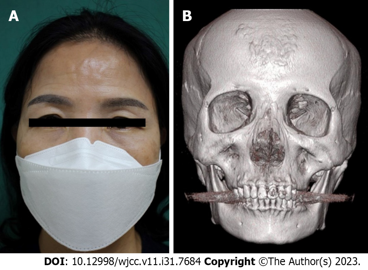
Figure 1 Preoperative images.
A: Preoperative clinical photography of a 54-year-old female patient with disseminated osteoma on the forehead; B: Preoperative computed tomography scan showing wide inoculation of osteoma in the frontal bone.
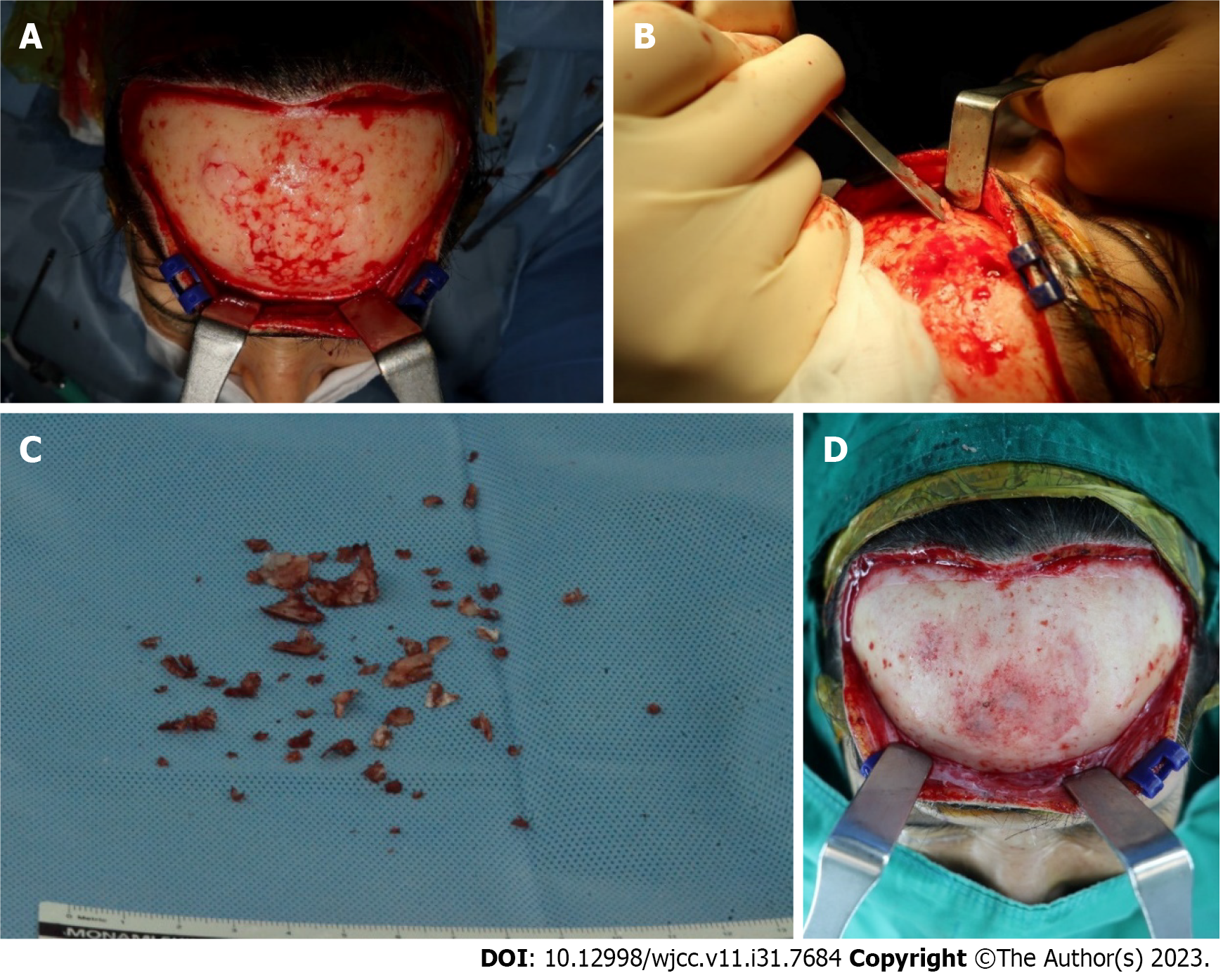
Figure 2 Intraoperative view.
A: Widely disseminated osteoma on the frontal bone; B: Ostectomy with osteotome and mallet; C: Resected bony particles; D: Bone wax application on the excised surface.
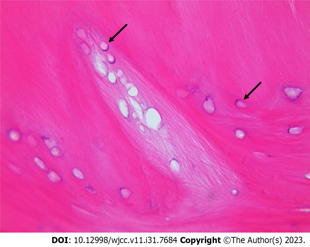
Figure 3 Pathological examination of the resected specimen revealed a benign osteoma of the compact bone type, without any cancellous tissue.
Multiple osteocytes (black arrow) were identified without active osteoblast (hematoxylin and eosin stain, × 400).
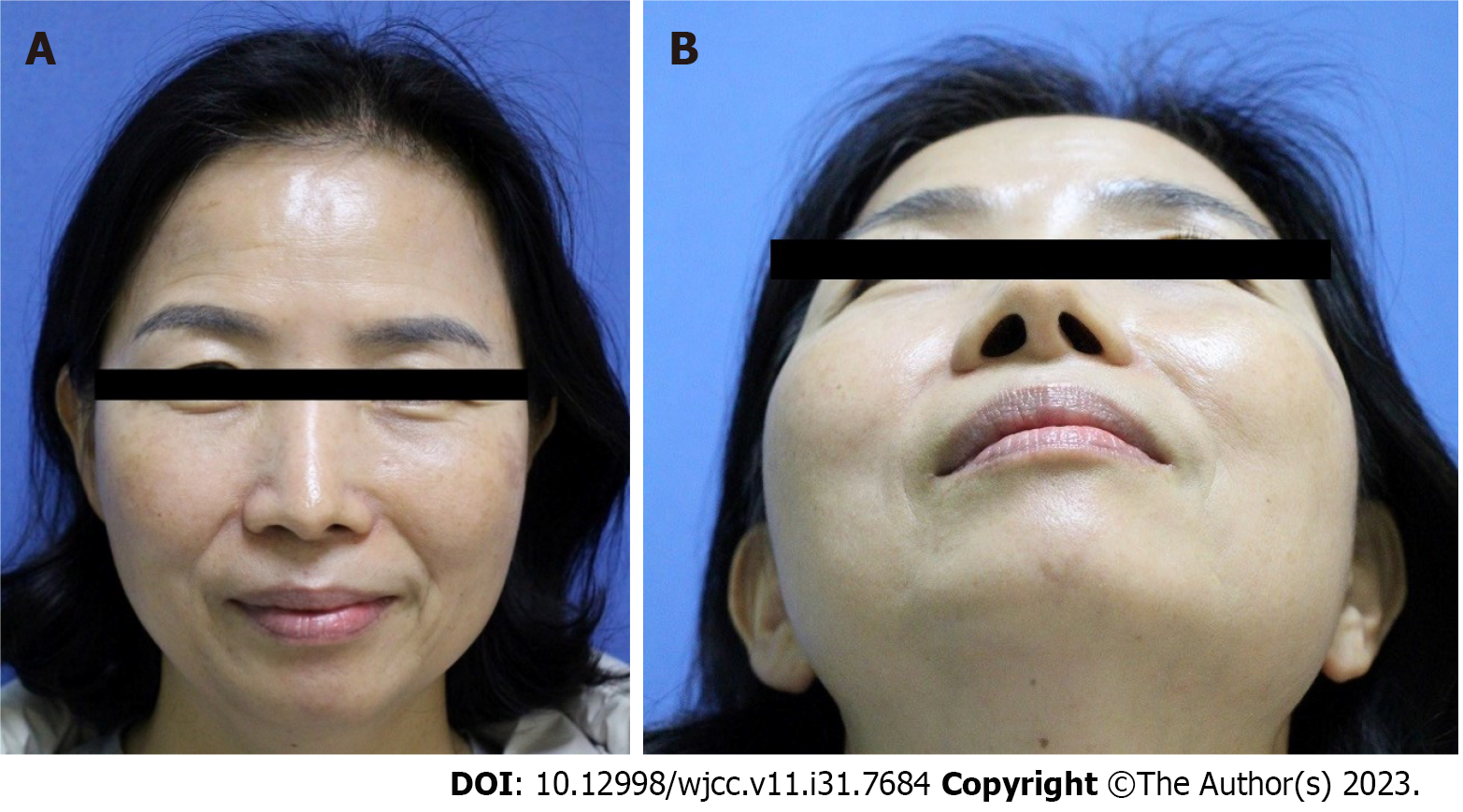
Figure 4 Clinical photograph in the sixth postoperative month.
A: Frontal view; B: Worm’s eye view.
- Citation: Lee DY, Lim S, Yoon JS, Eo S. Recurred forehead osteoma disseminated after previous osteoma excision: A case report. World J Clin Cases 2023; 11(31): 7684-7689
- URL: https://www.wjgnet.com/2307-8960/full/v11/i31/7684.htm
- DOI: https://dx.doi.org/10.12998/wjcc.v11.i31.7684


