Copyright
©The Author(s) 2023.
World J Clin Cases. Nov 6, 2023; 11(31): 7635-7639
Published online Nov 6, 2023. doi: 10.12998/wjcc.v11.i31.7635
Published online Nov 6, 2023. doi: 10.12998/wjcc.v11.i31.7635
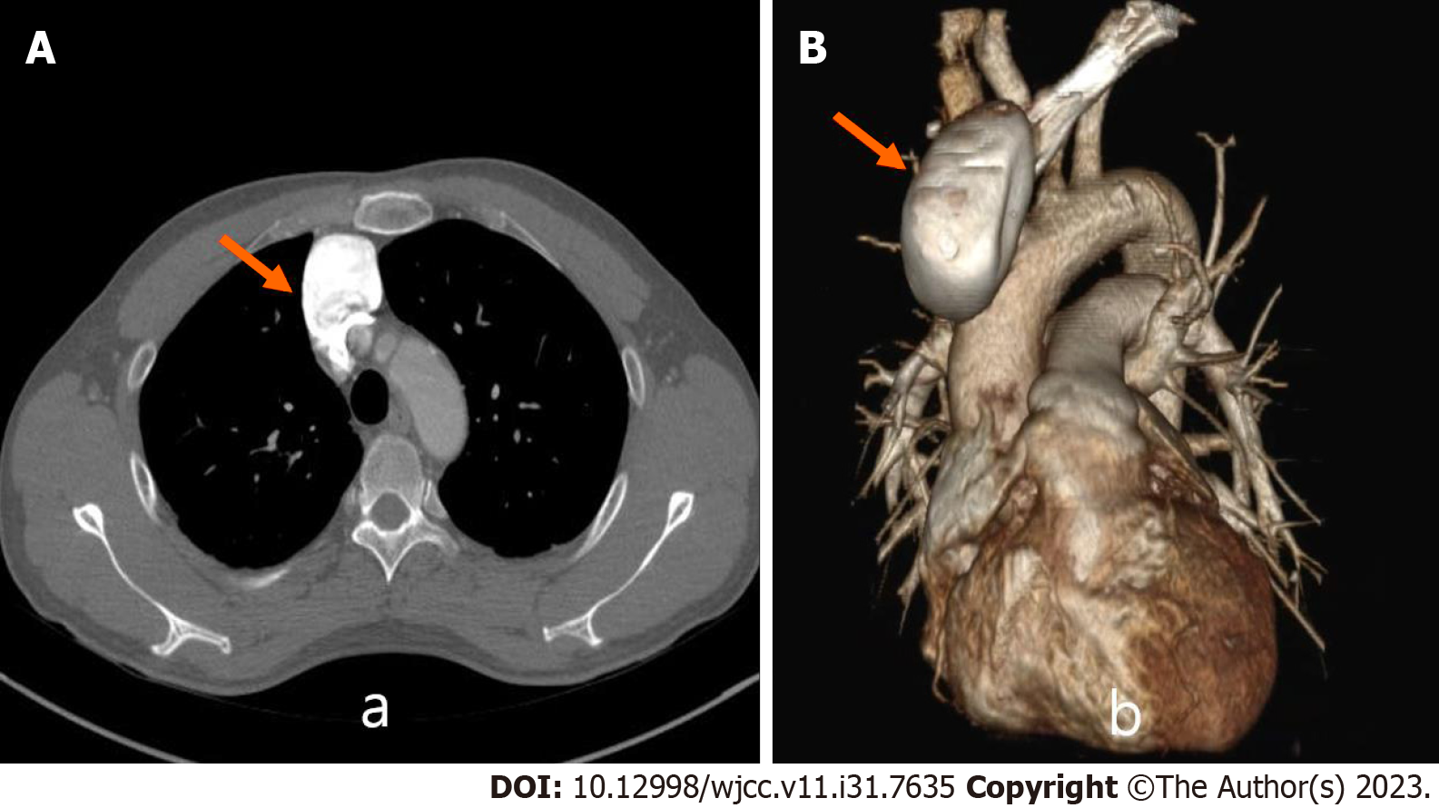
Figure 1 A chest computed tomography scan revealed a 6.
2 cm aneurysm in the left innominate vein and superior vena cava junction. A: Superior vena cava aneurysm of about 6.2 cm in size seen on chest computed tomography in the axial view; B: Three-dimensional reconstruction image.
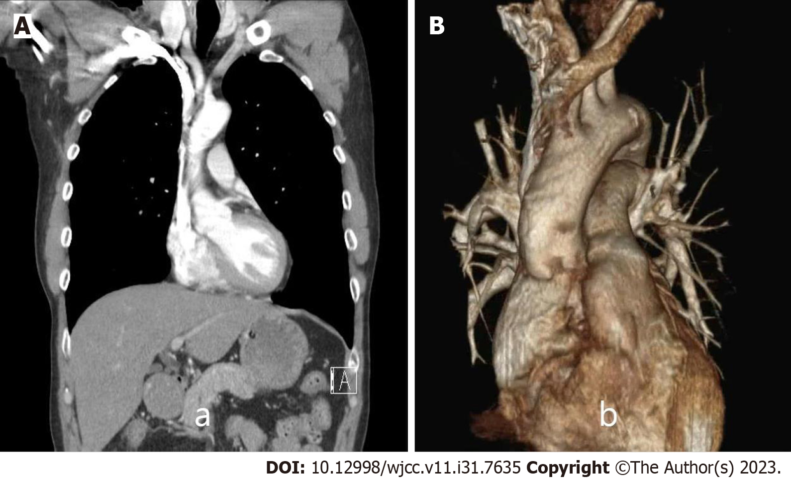
Figure 2 The patient was under follow-up, and no specific findings were observed on computed tomography 1 year after surgery.
A: Computed tomography image 1 year after surgery in the coronal view; B: Three-dimensional reconstruction image.
- Citation: Kim SP, Son J. Simultaneous lateral and subxiphoid access methods for safe and accurate resection of a superior vena cava aneurysm: A case report. World J Clin Cases 2023; 11(31): 7635-7639
- URL: https://www.wjgnet.com/2307-8960/full/v11/i31/7635.htm
- DOI: https://dx.doi.org/10.12998/wjcc.v11.i31.7635


