Copyright
©The Author(s) 2023.
World J Clin Cases. Oct 26, 2023; 11(30): 7393-7397
Published online Oct 26, 2023. doi: 10.12998/wjcc.v11.i30.7393
Published online Oct 26, 2023. doi: 10.12998/wjcc.v11.i30.7393
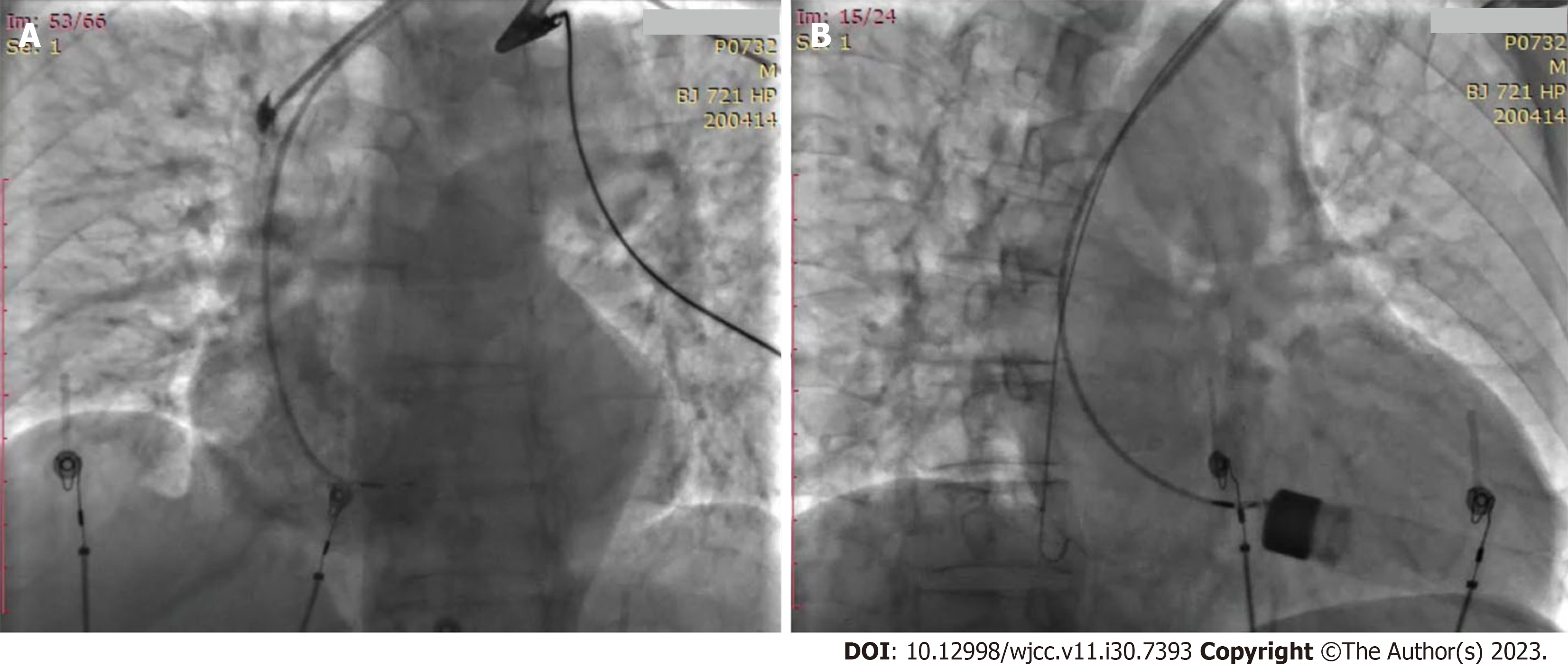
Figure 1 Right ventricular angiography showed the lead helix wound with the chordae tendineae.
A: LAO 30°; B: RAO 30°.
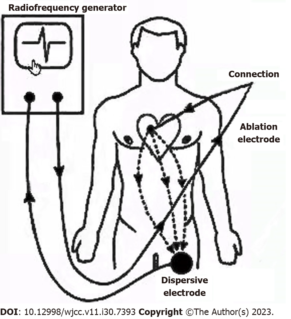
Figure 2 Loop schematic diagram of the chordae tendineae was ablated by radiofrequency electricity through the tip of the Model 3830 lead (radiofrequency generator-ablation catheter-3830 lead tip-chordae tendineae-blood-heart-back electrode plate-radiofrequency generator).
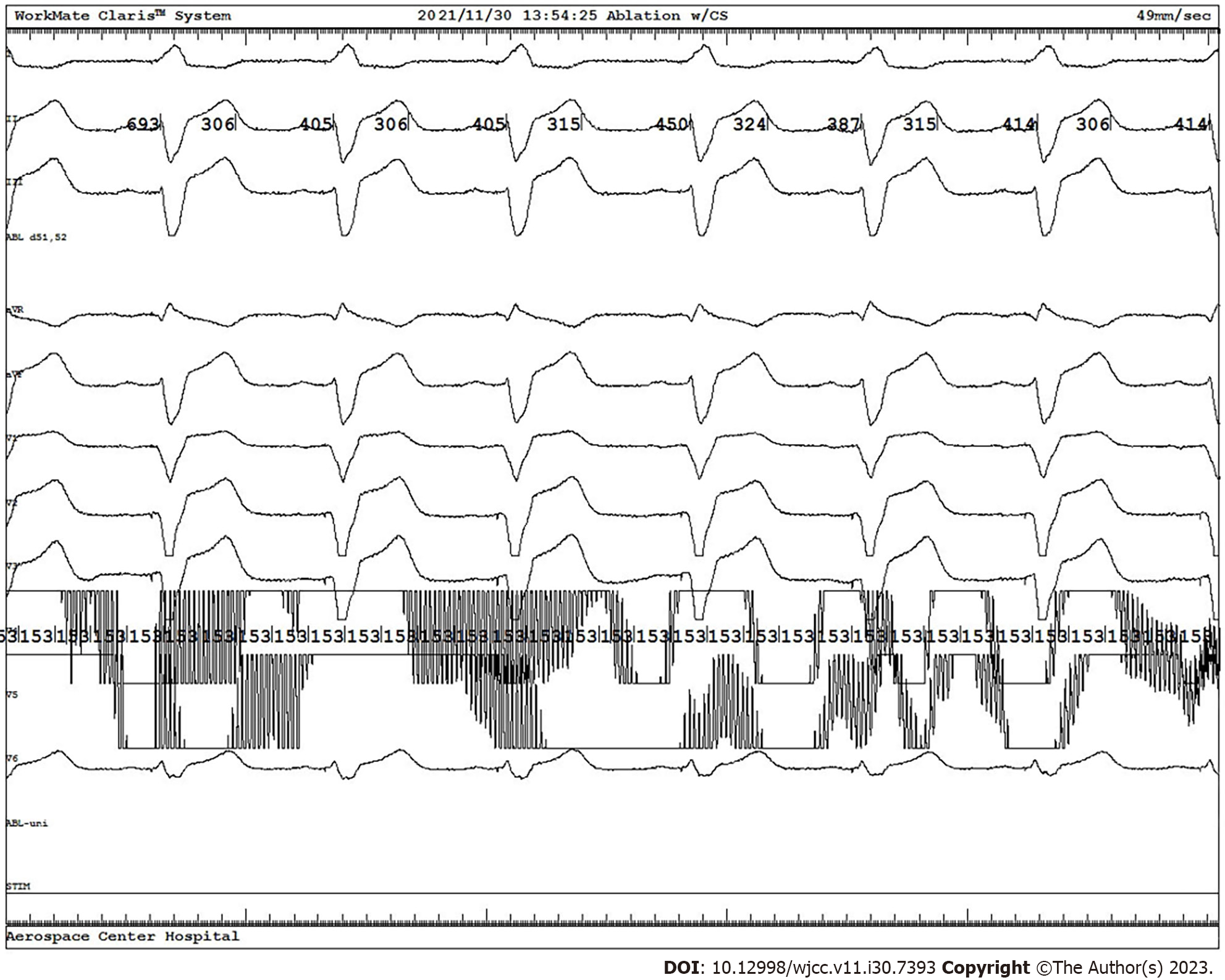
Figure 3 Intracardiac electrocardiogram of left bundle branch pacing.
- Citation: Liu TF, Ding CH. Lead helix winding tricuspid chordae tendineae: A case report. World J Clin Cases 2023; 11(30): 7393-7397
- URL: https://www.wjgnet.com/2307-8960/full/v11/i30/7393.htm
- DOI: https://dx.doi.org/10.12998/wjcc.v11.i30.7393


