Copyright
©The Author(s) 2023.
World J Clin Cases. Oct 26, 2023; 11(30): 7386-7392
Published online Oct 26, 2023. doi: 10.12998/wjcc.v11.i30.7386
Published online Oct 26, 2023. doi: 10.12998/wjcc.v11.i30.7386
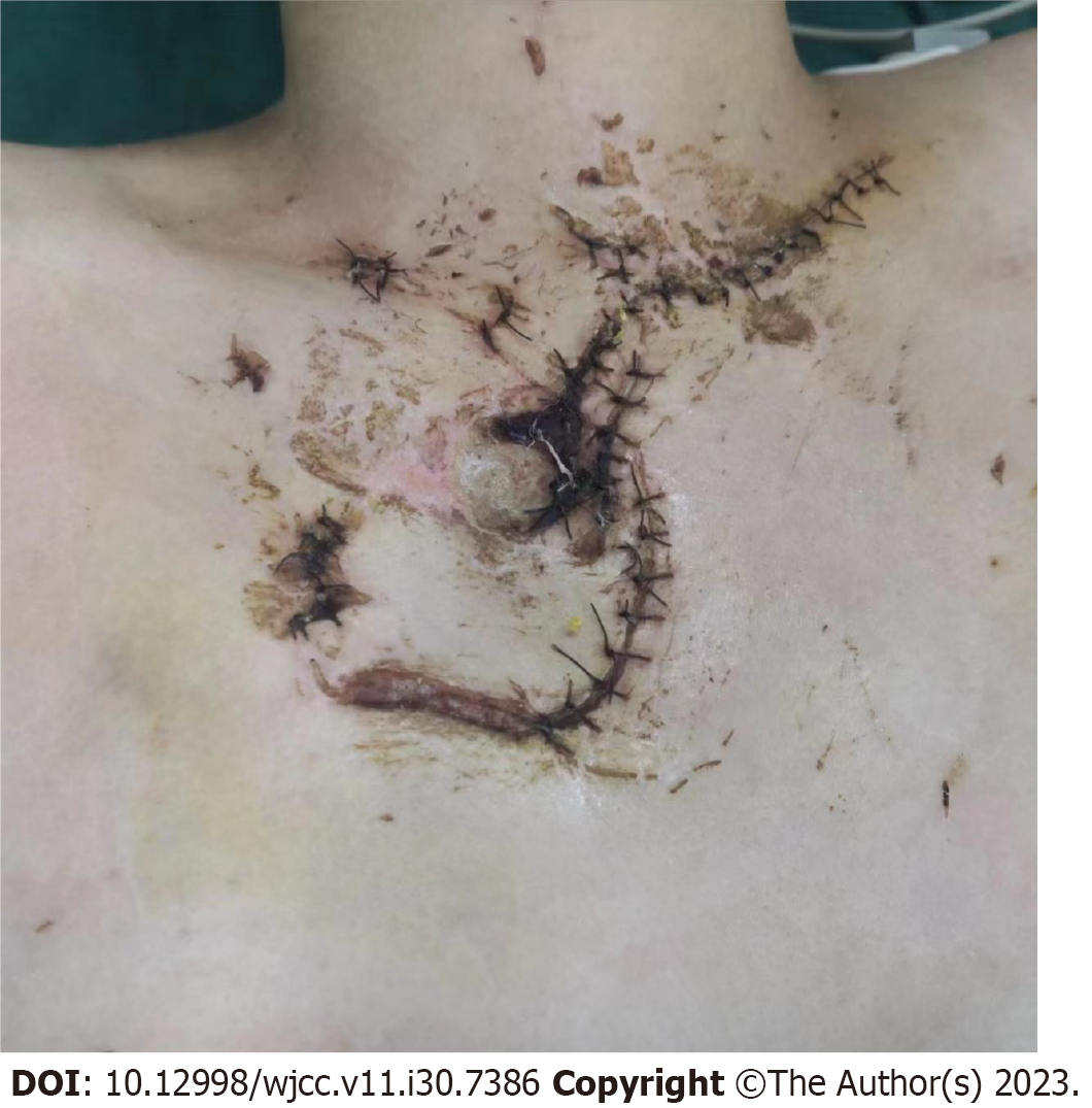
Figure 1 Necrosis and infection can be observed around the sutured irregular anterior chest wound.
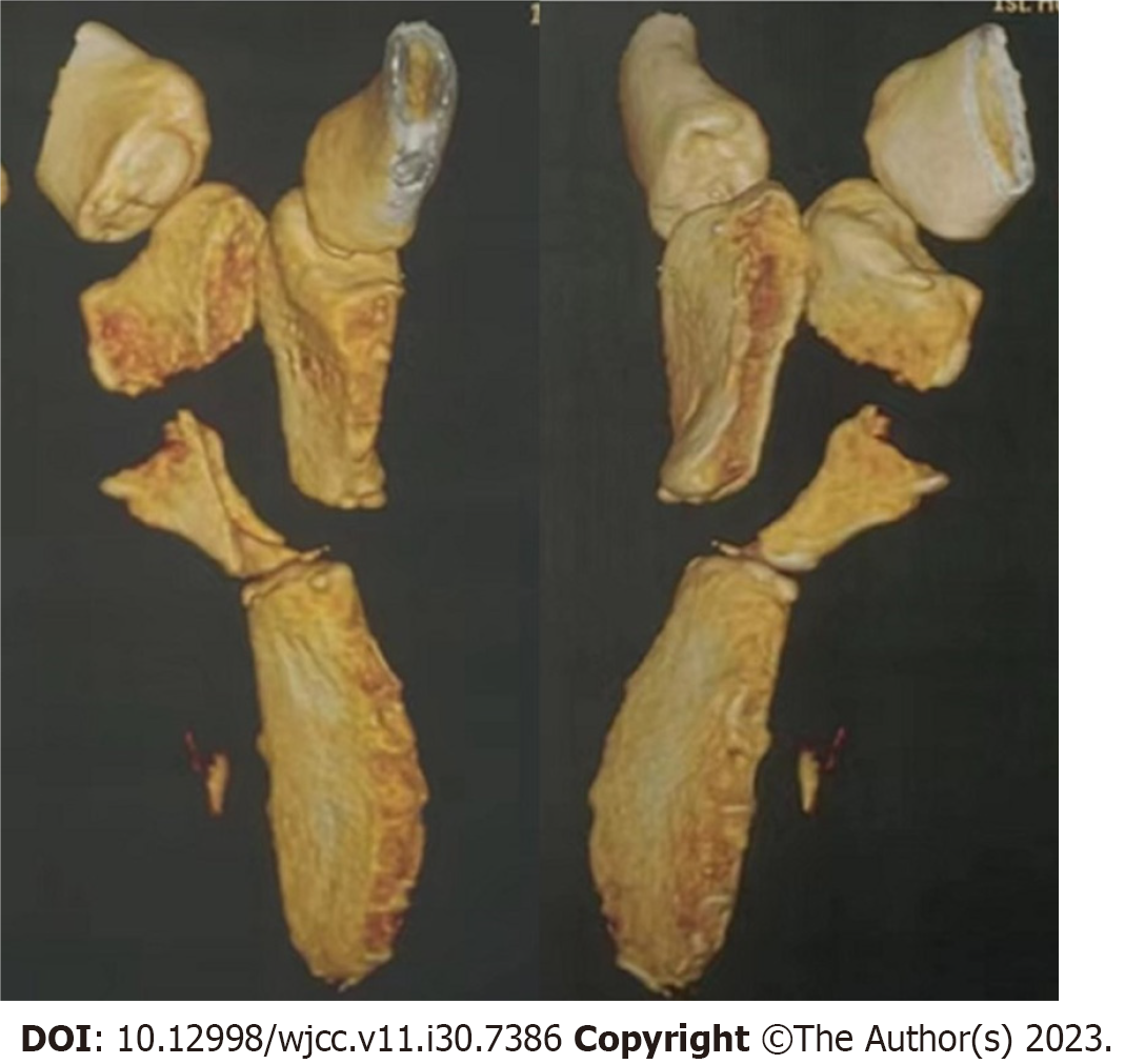
Figure 2 Computed tomographic three-dimensional reconstruction image of the sternum.
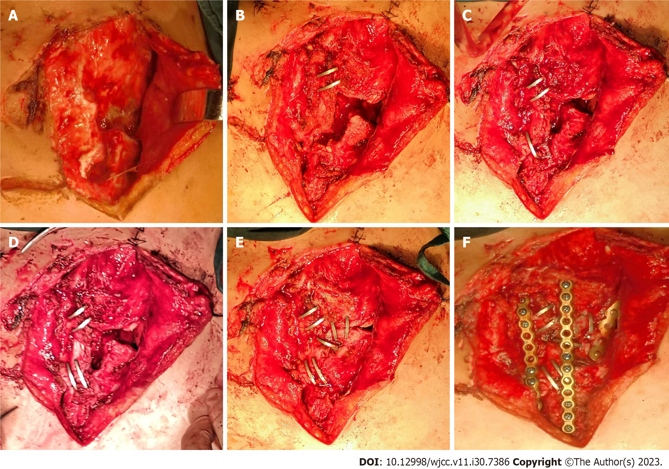
Figure 3 Images during the surgical procedure.
A: The crushed manubrium sterni was revealed after the removal of the sutures; B-E: Repositioning and primary fixation of the manubrium sterni fragments with the shape-memory alloy staples; F: Manubriosternal joint dislocation was reinforced and the left first rib cartilage fracture was fixed.
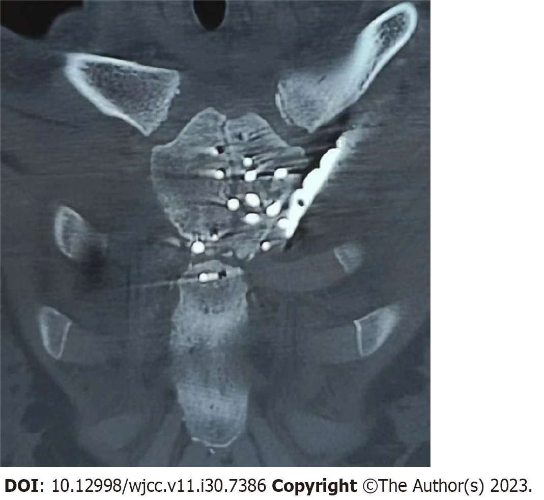
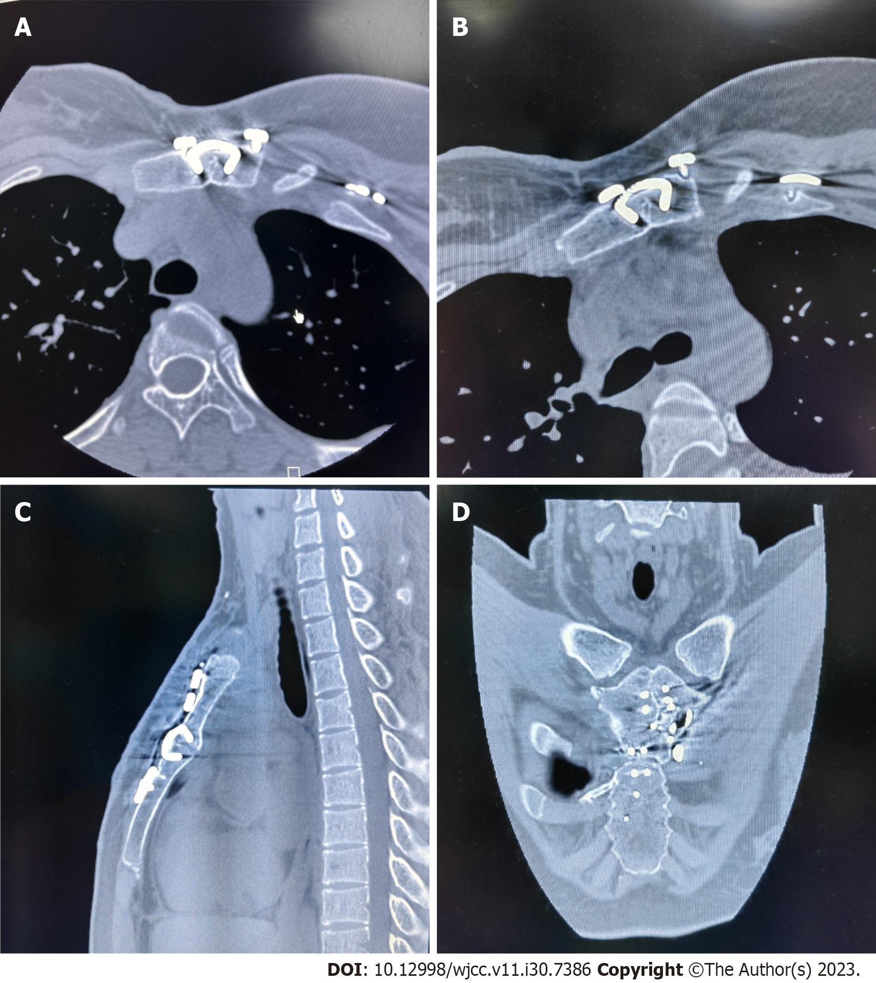
Figure 4 Computed tomography image of the sternum after operation.
Figure 5 Computed tomography image of the sternum at follow-up.
A-C: No staple loosening was observed; D: Fractures are healing well.
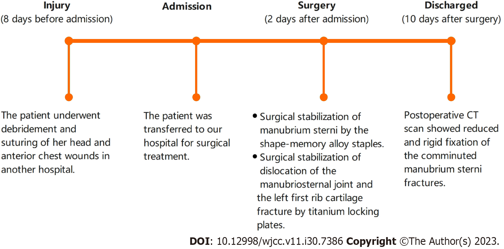
Figure 6 Timeline.
CT: Computed tomography.
- Citation: Zhang M, Jiang W, Wang ZX, Zhou ZM. Using shape-memory alloy staples to treat comminuted manubrium sterni fractures: A case report. World J Clin Cases 2023; 11(30): 7386-7392
- URL: https://www.wjgnet.com/2307-8960/full/v11/i30/7386.htm
- DOI: https://dx.doi.org/10.12998/wjcc.v11.i30.7386


