Copyright
©The Author(s) 2023.
World J Clin Cases. Jan 26, 2023; 11(3): 692-699
Published online Jan 26, 2023. doi: 10.12998/wjcc.v11.i3.692
Published online Jan 26, 2023. doi: 10.12998/wjcc.v11.i3.692
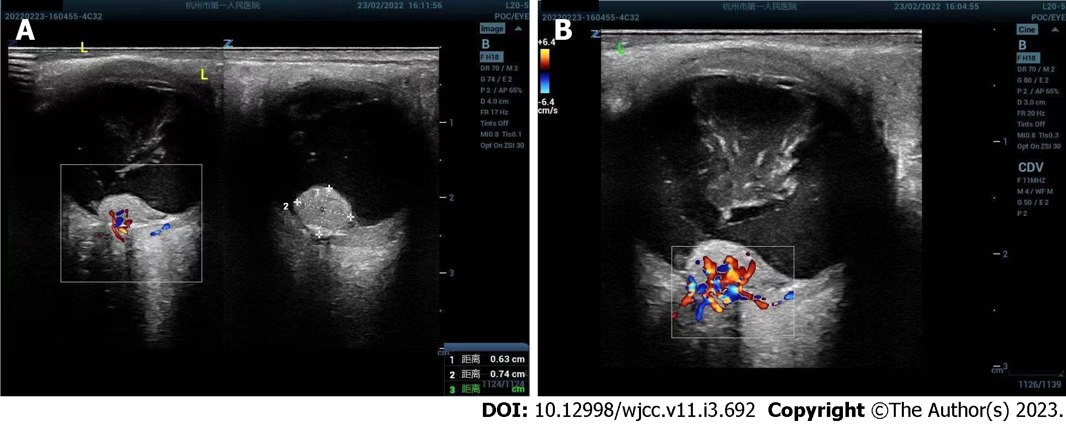
Figure 1 Ultrasound images of left retinal hemangioblastoma.
A: Ultrasound showed an irregular isoechoic mass of about 6.3 mm × 7.4 mm in front of the left optic nerve head; B: Color doppler flow imaging showed abundant blood flow signals in the lesion.
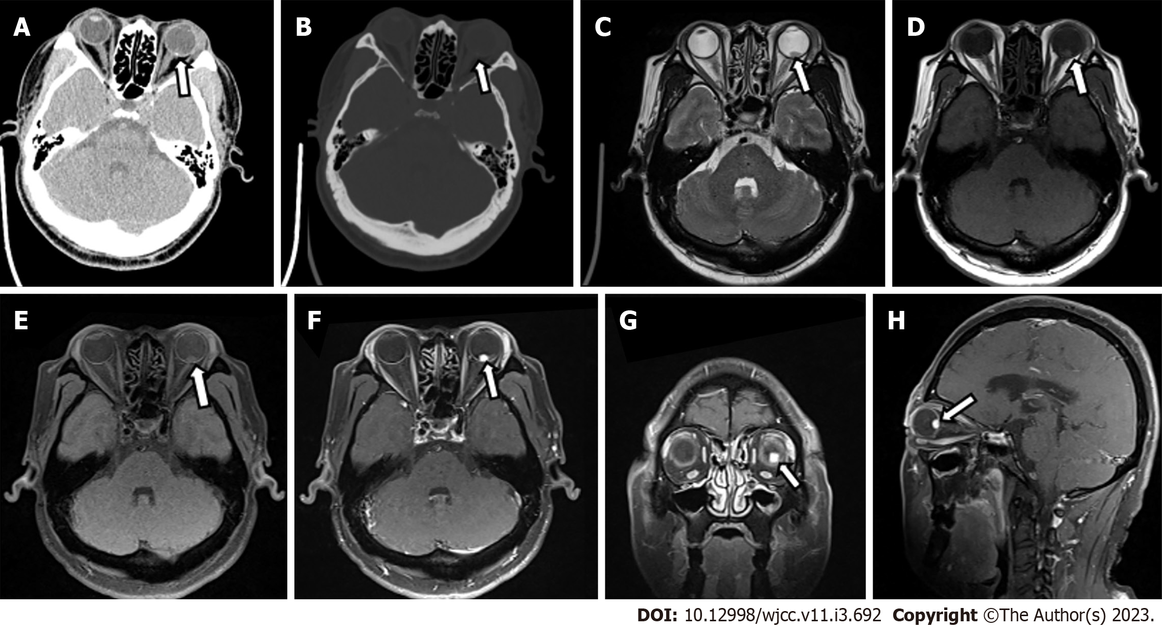
Figure 2 Computed tomography and magnetic resonance imaging of left retinal hemangioblastoma.
A: Computed tomography (CT) transverse soft tissue window of orbit showed punctate calcification on the posterior wall of the left eye ring and small patchy soft tissue density in the posterior part of the eyeball. The lesion measured about 5 mm × 8 mm, with an ill-defined border; B: CT transverse bone window of orbital showed no obvious abnormal change of orbital bone; C: The lesion was hypointense on transaxial T2-weighted sequence; D and E: The lesion was slightly hyperintense on transaxial T1-weighted images (D) and transaxial T1-weighted + fat-suppression images (E); F-H: Left posterior para-bulbar lesions were significantly enhanced on gadolinium-enhanced T1-weighted + fat-suppression images [mainly included transverse (F), coronal (G), and sagittal sequences (H)] (White arrows represent lesion).
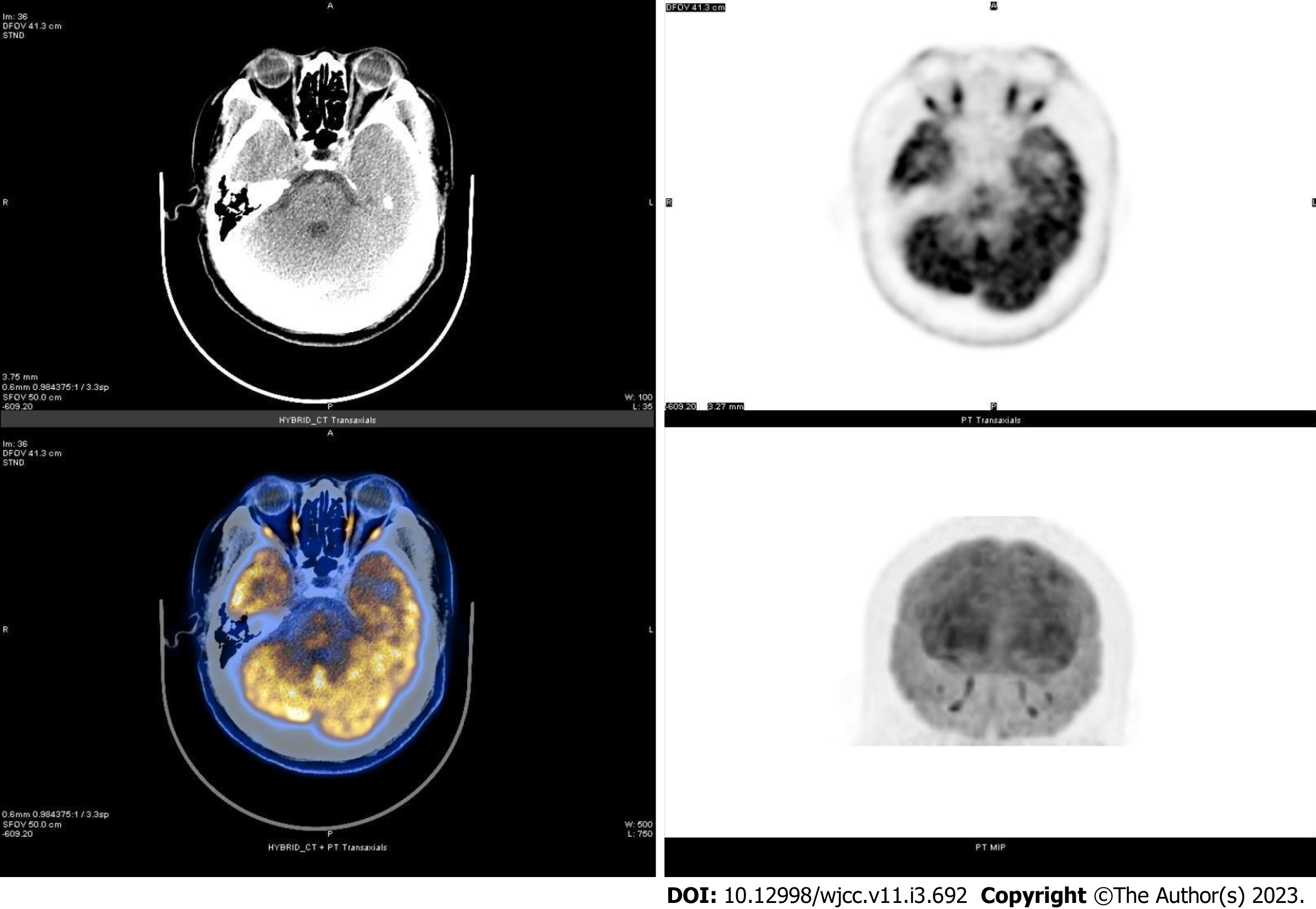
Figure 3 Transaxial computed tomography image, transaxial and coronal positron emission tomography metabolograms, color fusion map of positron emission tomography / computed tomography images at the orbital level.
The transaxial computed tomography (CT) image at the orbital level showed a patchy slightly hyperdense lesion. The transaxial positron emission tomography (PET) metabologram, coronal PET metabologram and PET/CT color fusion map at the orbital level showed no metabolic changes, and its SUVmax was 50.9.
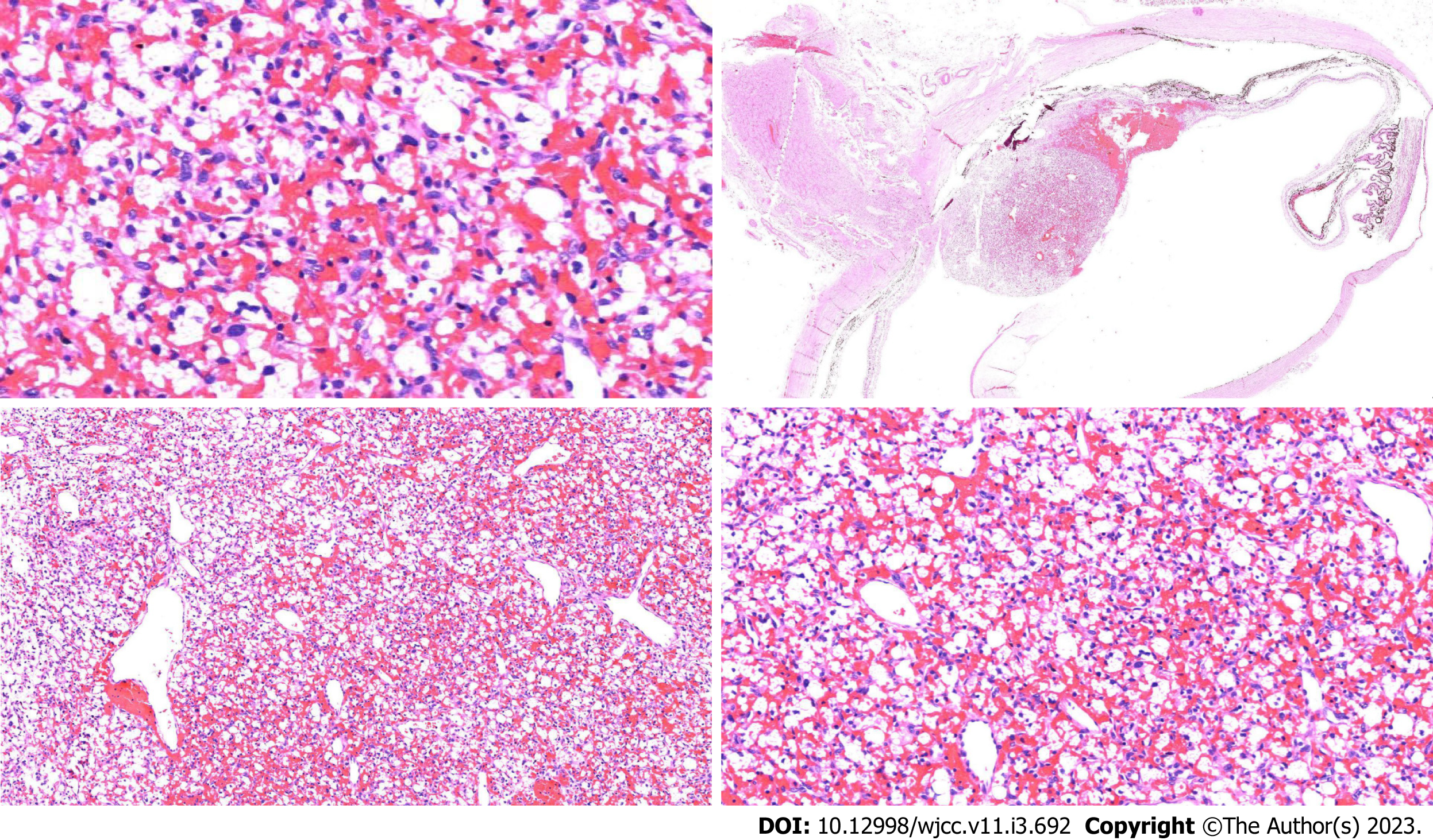
Figure 4 Postoperative histopathological and immunohistological images of left retinal hemangioblastoma.
The left eyeball lesions were mainly composed of two components, capillaries and interstitial cells surrounded by vacuolated or eosinophilic cytoplasm, which showed epithelioid stromal cells and staghorn dilated thin-walled vessels in capillaries (hematoxylin-eosin staining, magnification × 4).
- Citation: Tang X, Ji HM, Li WW, Ding ZX, Ye SL. Imaging features of retinal hemangioblastoma: A case report. World J Clin Cases 2023; 11(3): 692-699
- URL: https://www.wjgnet.com/2307-8960/full/v11/i3/692.htm
- DOI: https://dx.doi.org/10.12998/wjcc.v11.i3.692


