Copyright
©The Author(s) 2023.
World J Clin Cases. Jan 26, 2023; 11(3): 684-691
Published online Jan 26, 2023. doi: 10.12998/wjcc.v11.i3.684
Published online Jan 26, 2023. doi: 10.12998/wjcc.v11.i3.684
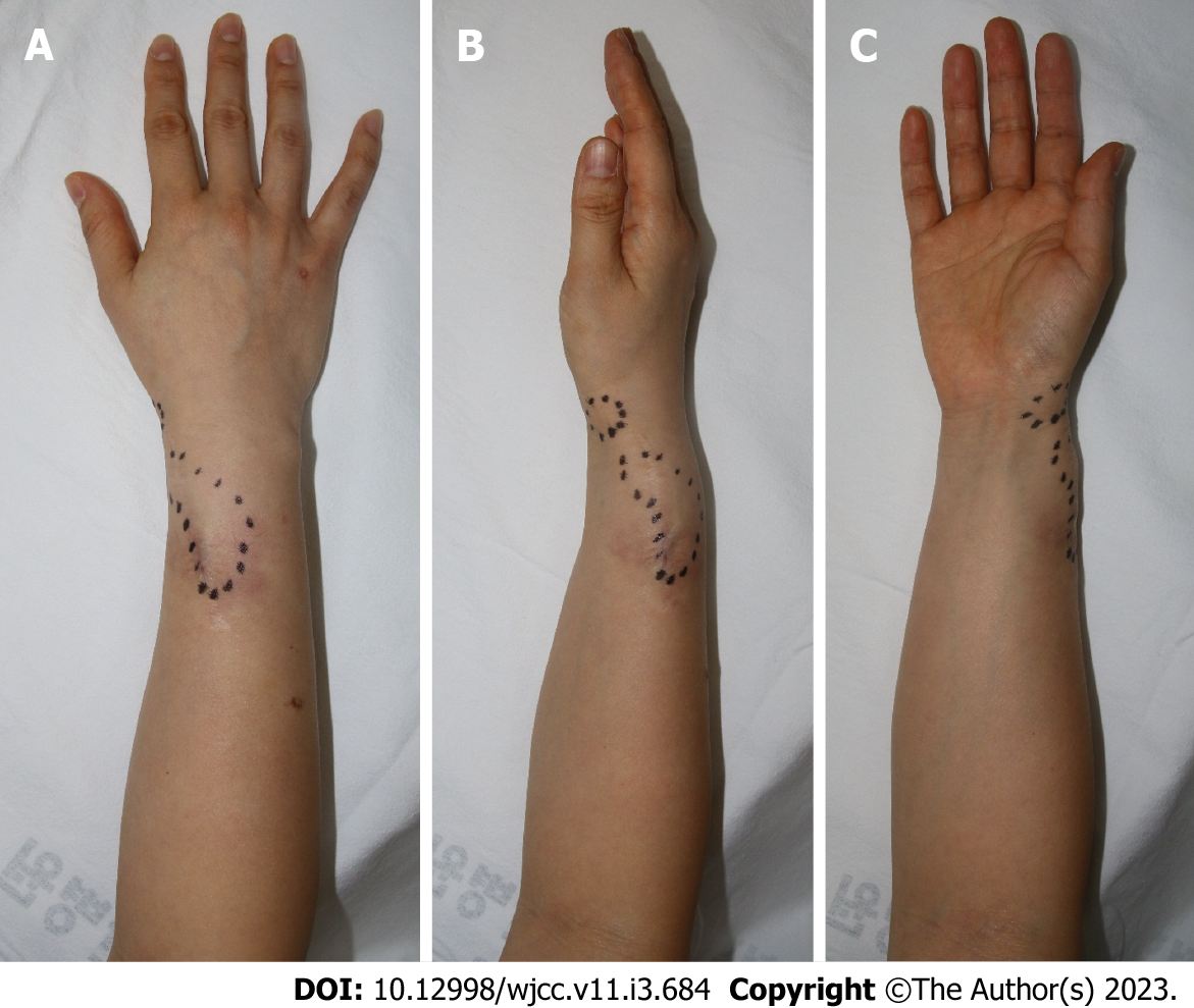
Figure 1 Preoperative photographs.
A: One 2 cm × 5 cm in size in the medial part of the forearm; B and C: A soft, painless lump was palpated along the extensor pollicis brevis of the right thumb, one 1 cm × 2 cm in size on the medial side of the right wrist. The black dotted line signifies the border of the lump to the touch. The patient’s sensation, range of motion of fingers and wrists, and motor power were normal. The previously operated scar is identified about 6 cm on the radial side of the right forearm.
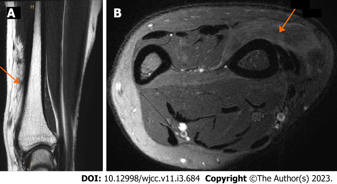
Figure 2 Preoperative magnetic resonance imaging showing lipomatous masses with fat-like attenuation in extensor pollicis brevis muscles and ligaments.
A: In the coronal view, lipomatous mass infiltrated in the muscle was identified. Boundaries with surrounding muscles and ligaments were unclear; B: In the axial view, lipomatous mass of similar attenuate to fat was identified. An orange arrow indicates a lipomatous mass that infiltrates muscle and ligaments.
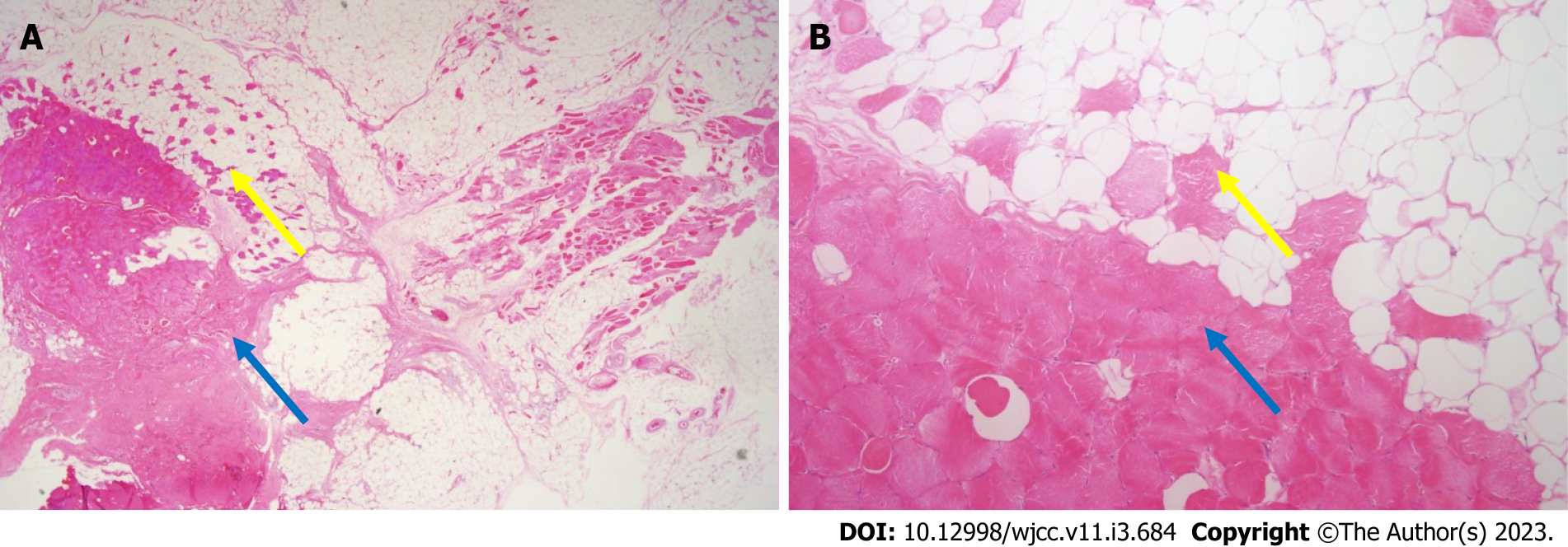
Figure 3 Histological findings.
A: Microscopic findings of intramuscular lipomas showing mature adipocytes and skeletal muscle fibers (pink color) (× 10.25 magnification, hematoxylin and eosin staining); B: Skeletal muscle fibers (pink color) are observed between mature adipocytes (× 100 magnification, hematoxylin and eosin staining). Yellow arrows indicate muscle components in fat. Blue arrows indicate muscle belly of extensor pollicis brevis.
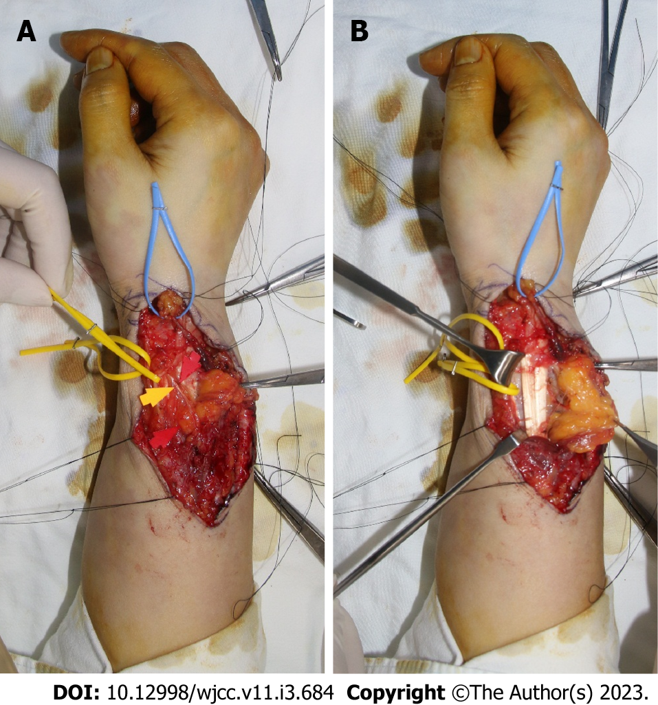
Figure 4 Intraoperative photographs.
A: The previous surgical scar was used, and an incision was applied along the run of the extensor pollicis brevis (EPB) to expose the lipomatous mass. A radial artery marked with yellow vessel loop and lipoma in the EPB tendon sheath marked with blue vessel loop were identified; B: Lipoma infiltrating into the muscle portion of EPB was removed while preserving the superficial radial nerve marked with yellow and red arrows.
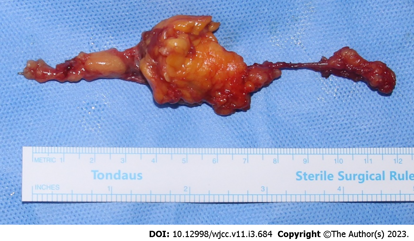
Figure 5 Photograph of the specimen.
A lipomatous mass of 12 cm × 4 cm × 5 cm was excised. It infiltrated the extensor pollicis brevis (EPB) muscle and tendon sheath. Most of the mass burden was located in the muscle portion of the EPB, presenting as yellow tissue with lobulated aspect and muscular fibers throughout.
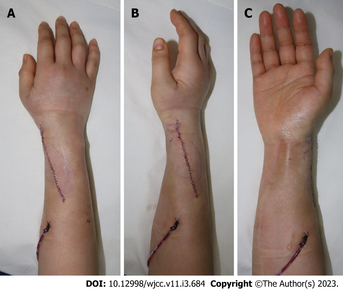
Figure 6 Postoperative photographs.
A and B: Primary closure was performed after removal of the intramuscular lipoma and 200cc Hemovac was inserted. There was no contour deformity on postoperative physical examination. Sensory function was intact, and circulation was well maintained; C: However, some extensor pollicis brevis motor weakness occurred. Nevertheless, no deformation of the thumb was seen in the neutral state. Other ranges of motion remained intact.
- Citation: Byeon JY, Hwang YS, Lee JH, Choi HJ. Recurrent intramuscular lipoma at extensor pollicis brevis: A case report. World J Clin Cases 2023; 11(3): 684-691
- URL: https://www.wjgnet.com/2307-8960/full/v11/i3/684.htm
- DOI: https://dx.doi.org/10.12998/wjcc.v11.i3.684


