Copyright
©The Author(s) 2023.
World J Clin Cases. Oct 16, 2023; 11(29): 7253-7257
Published online Oct 16, 2023. doi: 10.12998/wjcc.v11.i29.7253
Published online Oct 16, 2023. doi: 10.12998/wjcc.v11.i29.7253
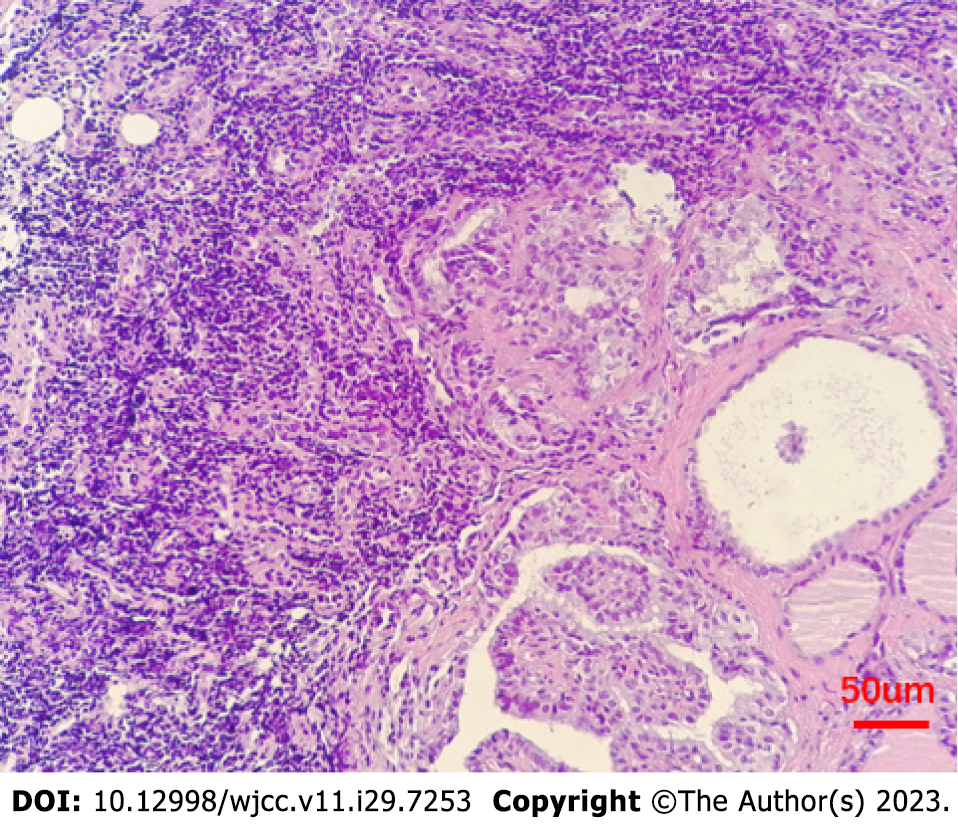
Figure 1 Pathological reveal: Metastatic carcinoma in lymphoid tissue, combined with morphology in line with thyroid papillary carcinoma metastasis (hematoxylin and eosin stain, 200 ×).
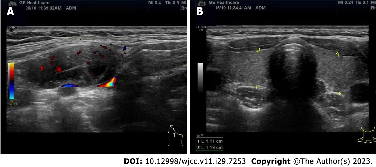
Figure 2 Ultrasonography reveal.
A: 2.0 cm × 1.1 cm solid-cystic mass with clear border and regular shape, and the separation can be seen inside; B: The thyroid gland has no abnormalities.
- Citation: Chen GY, Li T. Submandibular solid-cystic mass as the first and sole manifestation of occult thyroid papillary carcinoma: A case report. World J Clin Cases 2023; 11(29): 7253-7257
- URL: https://www.wjgnet.com/2307-8960/full/v11/i29/7253.htm
- DOI: https://dx.doi.org/10.12998/wjcc.v11.i29.7253


